
Application of Nanoparticle Drug Delivery Systems in the Medical Field: Focusing on Functionalized Gold Nanomaterials
Yanqi Wu1,2, Hui Chen3, Song Li3, Zhishan Yuan4, Yuhong Guan2,5, Nongyue He5,6*
1State Key Laboratory of Quality Research in Chinese Medicine, Macau University of Science and Technology, Macau, China
2Shenzhen LemnisCare Medical Technology Co., Ltd., Shenzhen, Guangdong Province, China
3Institute of Future Science, University of South China, Hengyang, Hunan Province, China
4School of Electromechanical Engineering, Guangdong University of Technology, Guangzhou, Guangdong Province, China
5Hunan Key Laboratory of Biomedical Nanomaterials and Devices, Hunan University of Technology, Zhuzhou, Hunan Province, China
6State Key Laboratory of Bioelectronics, National Demonstration Center for Experimental Biomedical Engineering Education, School of Biological Science and Medical Engineering, Southeast University, Nanjing, Jiangsu Province, China
*Correspondence to: Nongyue He, PhD, Professor, Hunan Key Laboratory of Biomedical Nanomaterials and Devices, Hunan University of Technology, No. 218 Daping Road, Zhuzhou 412007, Hunan Province, China; State Key Laboratory of Bioelectronics, National Demonstration Center for Experimental Biomedical Engineering Education, School of Biological Science and Medical Engineering, Southeast University, No. 2 Southeast University Road, Nanjing 210096, Jiangsu Province, China; Email: nyhe@seu.edu.cn
DOI: 10.53964/jmn.2023005
Abstract
Nanoparticle drug delivery systems are currently one of the hottest topics in the field of medicine. They can precisely deliver drugs to the parts of the patient that need treatment, with the goal of improving patient effectiveness and reducing drug toxicity. Gold nanoparticles (AuNPs) are considered one of the most promising nanomaterials due to their unique optical, electronic, sensing, and biochemical properties. By combining AuNPs with different ligands, functionalized gold nanoparticle (F-AuNPs) drug delivery systems can be obtained, which have broad application prospects in the field of medicine. This article focuses on the different ligand connection types of F-AuNPs and reviews the research progress of F-AuNPs drug delivery systems in the field of medicine in recent years.
Keywords: medicine, nanoparticle drug delivery system, functionalization, gold nanoparticles, review
1 INTRODUCTION
Nanomedicine belongs to the field of nanoscience, and the application of nanotechnology in the field of medicine has become a current research hotspot[1]. The development of nanoscience and the preparation of nanomaterials provide the foundation and technical support for the development of nanomedicine. The application of nanotechnology in medicine mainly includes nanosensors, nanomolecular imaging, nanodrug delivery and controlled release systems, and the application of nanomaterials in tissue engineering and regenerative medicine[2]. Nanomaterials have unique physical, chemical, and biological properties, such as a large specific surface area, strong controllability, and good biocompatibility, which provide an excellent platform for the development of nanomedicine[3]. By regulating the properties and structures of nanomaterials, precise transport and controlled release of drugs can be achieved, thus improving drug efficacy and reducing side effects[4]. In addition, the application of nanotechnology in medicine can also achieve precise diagnosis, personalized treatment, tissue repair, and other functions, which have a significant impact on human health[5].
Gold nanoparticles (AuNPs) are nanoparticles composed of gold atoms and have many unique physical, chemical, and biological properties[6]. These properties make AuNPs widely applicable in medicine, biology, chemistry, and materials science[7]. However, naked AuNPs are not stable and tend to aggregate and precipitate into metallic particles. Functionalized gold nanoparticles (F-AuNPs) are nanomaterials obtained by modifying the surface or interior of AuNPs with different ligands or functional molecules[8]. By modifying the surface or interior of F-AuNPs, the physical and chemical properties of AuNPs can be adjusted to meet specific application requirements[9]. Therefore, the application prospects of F-AuNPs in drug delivery are wider than those of naked AuNPs. This article reviews the research progress of F-AuNPs in drug delivery systems in the field of medicine in recent years, aiming to provide a reference for further development and application of F-AuNPs drug delivery systems.
2 INTRODUCTION TO F-AUNPS AND THEIR APPLICATIONS
F-AuNPs refer to AuNPs that have been surface-modified by connecting different molecules to their surface, giving them specific properties and functions[10]. These molecules can be biological molecules, fluorescent dyes, peptides, polymers, and so on. F-AuNPs have excellent optical, electronic, biosensing, and drug delivery properties, making them widely used in the fields of biomedical, optical, and electronic sciences. For example, in the biomedical field, F-AuNPs can be used for molecular diagnosis, photothermal therapy, gene delivery, and other applications[11]. In addition, F-AuNPs have good biocompatibility and low toxicity, making them a promising potential candidate for medical applications[12].
The conjugation strategy involves chemically reacting molecules with the chemical functional groups on the surface of AuNPs, forming a stable conjugated system. The conjugation strategy can be used to combine different molecules with AuNPs, forming F-AuNPs. In the biomedical field, the conjugation strategy is widely used for drug delivery, molecular diagnosis, and biological imaging (common F-AuNPs conjugation strategies are shown in Figure 1[13]).
|
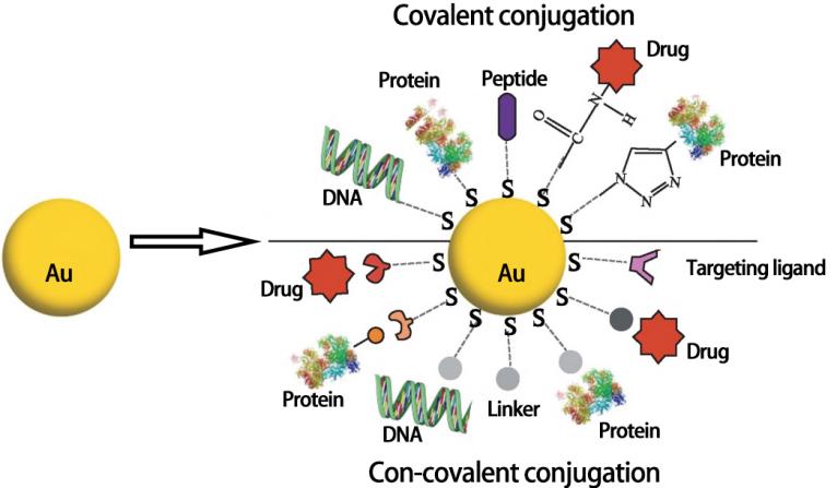
Figure 1. Common conjugation strategies for functionalized gold nanoparticles.
The purpose of functionalizing and modifying AuNPs is to give them more special properties and functions for use in different fields. The specific goals of functionalizing and modifying AuNPs include: (1) improving dispersibility and stability, which is achieved by conjugating molecules to the surface of AuNPs and thus preventing aggregation and precipitation; (2) changing optical properties, as AuNPs have unique optical properties that can be adjusted through surface functionalization, such as regulating the wavelength, intensity, and direction of absorption and scattering of light, for use in optical imaging and therapy; (3) increasing biocompatibility, which is realized by modifying molecules on the surface of AuNPs to reduce toxicity for use in the biomedical field; (4) increasing targeting, which is achieved by modifying targeting ligands on the surface of AuNPs to target specific cells, tissues, or biological molecules, for precision diagnosis and therapy; (5) enhancing drug delivery ability by modifying drugs on the surface of AuNPs, which can also improve therapeutic efficacy, and reduce drug side effects. In summary, the purpose of functionalizing and modifying AuNPs is to make them more applicable in different fields and realize their potential for various applications.
3 DIFFERENT LIGAND CONNECTION TYPES FOR F-AUNPS
3.1 Physical Adsorption Connection Type
The physical adsorption connection type is a method of adsorbing molecules or compounds onto the surface of AuNPs, which does not involve the formation of covalent bonds[14]. The commonly used physical adsorption connection types include electrostatic interaction, van der Waals forces, and hydrophobic interaction. As its connection strength is usually weak and easily influenced by environmental factors, the simplest and fastest ligand connection method is not suitable for applications that require high connection stability[15]. However, research[16] has shown that the dissociation constant (pKa) value of the ligand, the pH value of the surrounding solution, and the ion strength will all affect the size of the surface charge, thereby affecting the repulsion strength and stability of the AuNPs. Therefore, it is speculated that in the process of using AuNPs, their stability can be ensured by appropriately adjusting the pH value and ion strength of the ligand. Andrade et al.[17] used cetuximab to coat AuNPs, and the resulting nanocomposites maintained a stable level of tolerance for up to 2 years. Moreover, it effectively promoted the binding of epidermal growth factor receptors and induced tumor cell apoptosis. Khodashenas et al.[18] combined gelatin with AuNPs and used it as a drug carrier for curcumin. They studied the release curve of curcumin at different pH values (7.4 and 5.4) for 48h at 37℃, and the results showed that the amount of released drug under pH 5.4 conditions was larger than that under pH 7.4 conditions, and the release rate was slower. This study confirmed that physical adsorption of gelatin and AuNPs is more stable under acidic conditions. Although the physical adsorption ligand connection method is simple and easy to implement, it is not perfect, and fixing the ligand by physical adsorption can lead to random ligand connections. Some active sites may be blocked by adjacent biomolecules or AuNPs, so only a small fraction of the ligands may be active in the end.
3.2 Coordination Self-assembly Connection Type
Coordination self-assembly connection type is a method of connecting organic molecules containing thiol (-SH) with AuNPs[19]. Thiol has a strong affinity for gold and can form strong coordination bonds with gold atoms on the surface, thus connecting AuNPs with organic molecules[20]. Sulfide (RS-) is the most commonly used organic ligand, which can form very stable chemical bonds by coordinating with gold atoms on the surface. In addition, phosphate groups, amino groups, carboxyl groups, and other groups can also coordinate with gold atoms on the surface to achieve functional modification of AuNPs[21]. This connection method has high stability, but the number of connections is limited, and the molecular structure and atomic properties of the gold surface need to be considered during the connection, which requires higher experimental conditions. Bessar et al.[22] used 3-MPS containing thiol group to bind AuNPs to prepare water-soluble F-AuNPs and loaded them with MTX to treat psoriasis. The results showed that the drug loading rate of Au-3MPS could reach 70%-80%, and the release rate within 24h could reach 95%. In vitro studies have shown that compared with simple MTX, the use of Au-3MPS@MTX conjugates can further increase the delivery of MTX in the epidermis and dermis, and no skin retention of Au-3MPS was observed, which confirms that Au-3MPS@MTX conjugates can be used as carriers for treating local psoriasis.
Polymers are high molecular weight compounds formed by connecting many monomer molecules through covalent or ionic bonds[23]. In various fields such as chemistry, materials science, and biology, polymers have been widely used in the preparation of materials, drugs, biosensors, and more. In the preparation of nanoparticles, polymers can act as protective agents, controlling the size, shape, and surface properties of the nanoparticles[24]. Additionally, polymers can serve as carriers to encapsulate biological active substances such as drugs on the surface of nanoparticles, achieving precise delivery and release of drugs[25]. Therefore, polymers play an important role in the preparation and application of nanoparticles. In the field of AuNPs, many polymers can alter their size, shape, and stability. For example, the bifunctional linker of PEG is often used for surface modification of AuNPs[26]. It has been found that the orientation and density of ligands are two key factors that affect the surface properties of AuNPs[27]. Comenge and Puntes[28] found that when the density of the PEG-SH polymer, which was used to conjugate antitumor drug ligands onto the surface of AuNPs, was low, the PEG-SH polymer could bind to the AuNPs through multiple weak bonds. However, when the density of ligands was high, the surface was saturated with more stable Au-SH bonds. In contrast, Al-Harbi et al.[29] conducted a rat study by comparing blank AuNPs with PEG-coated AuNPs. The results showed that the PEG-coated AuNPs significantly increased the expression of pro-inflammatory cytokines in rat liver and kidney cells, confirming that the PEG-coated AuNP drug delivery system can further enhance the therapeutic effect of drugs compared to blank AuNPs.
3.3 Covalent Bonding Connection Type
Covalent bonding is another commonly used connection type for functionalizing AuNPs. Compared with physical adsorption and coordination self-assembly connection types, covalent bonding can provide more stable chemical bond connections and has greater flexibility in chemical modification[30]. Chemical modifiers commonly used on the surface of AuNPs include thiols, amines, and carboxylic acids, which can covalently bond with reactive groups on the gold surface, such as thiol groups, amino groups, and carbonyl groups[31]. In covalent bonding connection type, chemical modification usually requires certain reaction conditions and chemical catalysts, so it is necessary to strictly control the conditions and parameters of the reaction to avoid unnecessary side reactions or damage to the stability of the nanoparticles.Ghorbani and Hamishehkar[32] prepared polymer-coated AuNPs by covalently bonding MTX carboxylic acid with the amino group of the polymer shell of the AuNPs, thus developing a new pH-responsive nano platform for controlled and targeted delivery of anticancer drugs.
Magro et al.[33] has found that the size of AuNPs is similar to that of biological molecules such as proteins and DNA. Most peptides or proteins contain cysteine with thiol groups, which can be fixed on F-AuNPs through covalent bonding to exert their effects. Kalishwaralal et al.[34] found that PDCs can target and kill different types of cancer cells, but they are easily hydrolyzed in physiological environments. The study showed that the stability of PDCs can be improved by coupling them with polyethylene-coated AuNPs. In addition, the study also found that the coupling efficiency of PDCs and AuNPs may vary depending on the chemical properties of the drug and the specific amino acid sequence of the peptide, especially in highly hydrophobic conditions. Gasparri et al.[35] studied the effect of AuNPs coupled with head-tail cyclic peptide CGisoDGRG (Iso1) on mouse IL-12. The results showed that this AuNP conjugate was an effective strategy to improve its therapeutic index and maintain adoptive T cell therapy. Sun et al.[36] found that the conjugate (Au@DAPT@BSA) of F-AuNPs with BSA and 4,6-DAPT can reduce the minimum inhibitory concentration of gram-negative bacteria and broaden the spectrum of antibacterial activity against gram-positive bacteria, significantly reducing the number of Pseudomonas aeruginosa and Staphylococcus aureus in biofilms. In addition, Au@DAPT@BSA has a healing effect on subcutaneous abscesses caused by clinically isolated multidrug-resistant Escherichia coli or Staphylococcus aureus, without producing detectable toxicity. The above research results provide new strategies for improving the biocompatibility of nanomaterials with antibacterial drugs.
4 RESEARCH PROGRESS OF FUNCTIONALIZED GOLD NANOCARRIER SYSTEMS IN THE FIELD OF MEDICINE
4.1 Gene Therapy
In recent years, nanocarrier systems have been widely studied and applied in the field of medicine. In gene therapy, the application of functionalized gold nanocarrier systems has received great attention. Gene therapy is a technique that uses genetic engineering to introduce therapeutic genes into a patient’s body to treat diseases[37]. However, traditional gene therapy methods have many problems, such as low gene expression efficiency and poor safety, due to factors such as the instability of genes and difficulty in crossing the cell membrane. The unique properties of F-AuNPs, such as high surface area-to-volume ratio, good biocompatibility, and easy surface modification, make them an effective gene delivery platform[38]. To date, several functionalized gold nanocarrier systems have been designed for gene therapy, such as DNA-modified gold nanomaterials and siRNA-modified gold nanomaterials. In Ahwazi et al.’s study[39], researchers used a siRNA delivery system based on human immunodeficiency virus type 1 trans-activating transcriptional activator (HIV-1TAT) peptide-coated AuNPs to treat breast cancer. Positively charged nanoparticles have high cellular uptake ability, allowing for efficient gene transfection using low concentrations of nanoparticles. After transfection, ROR1 and its target gene CCND1 were downregulated, inducing apoptosis in cancer cells. This study demonstrated that HIV-1TAT peptide can bind to AuNPs via Au-S bonds and can be used as a carrier for negatively charged therapeutic agents for specific genes. In another study by Shaat et al.[40], to overcome the shortcomings of rapid enzymatic degradation and low transfection efficiency of siRNA, AuNPs modified with branched polyethyleneimine (bPEI) were used as intracellular siRNA delivery vehicles. The study showed that the complex enhanced the uptake of siRNA by cells without causing significant cytotoxicity. Moreover, the study confirmed that bPEI-modified AuNPs can be used as a safe carrier for delivering siRNA.
4.2 Targeted Drug Delivery
The application of functionalized gold nanocarriers in targeted drug delivery is also one of the most highly researched directions. Due to their excellent biological properties, F-AuNPs are very suitable as non-toxic carriers for targeted disease therapy[41]. The nanosize of F-AuNPs improves their circulation time, reduces aggregation rate, enhances the attachment to therapeutic molecules and target reagents, and further improves their ability to penetrate cell membranes while reducing overall cellular toxicity[42]. Therefore, F-AuNPs serve as effective means of targeted drug delivery.
4.2.1 Active Targeting
F-AuNPs have attracted attention as an effective means of drug delivery through active targeting, which can deliver drugs directly to target cells or tissues, thereby improving therapeutic efficacy and reducing unnecessary side effects. In active targeting, nanoparticles are conjugated to ligands that specifically bind to molecules that overexpress on the surface of target cells, but express poorly or do not exist at all in normal cells[43]. This ligand-receptor interaction induces nanoparticle internalization and drug release inside the cells through receptor-mediated endocytosis. Common ligands include antibodies, carbohydrates, enzymes, folate, peptides, and vitamins. Rizk et al.[44] conjugated AuNPs with TGF-β1 antibody, MTX, and tumor-targeting molecule folate. ELISA results showed that TGF-β1 antibody conjugated with AuNPs reduced extracellular TGF-β1 by 30% compared to the control group. F-AuNPs effectively reduced extracellular TGF-β1 levels, and folate and MTX enhanced toxicity against cancer cells. Thambiraj et al.[45] conjugated AuNPs with docetaxel in a non-covalent manner and linked them to the targeting molecule folate, and evaluated the cytotoxicity of the synthesized AuNP conjugates against lung cancer cell line H520. The results showed that AuNP-based nanocarriers could be used as a delivery system for various cancer chemotherapy drugs.
4.2.2 Passive Targeting
Passive targeting refers to the use of the unique physiological and pathological characteristics of tumor tissue to achieve the passive targeted delivery of drugs to tumor tissue by changing the size, shape, surface modification, and composition of nanoparticles in F-AuNP drug delivery systems[46]. A common passive targeting delivery strategy is to utilize the tumor tissue-specific EPR effect, that is, the high permeability of the vascular wall and the incomplete lymphatic drainage system in tumor tissue, allowing nanoparticles to enter tumor tissue through the vascular wall, and slow excretion of nanoparticles in tumor tissue, achieving passive targeted delivery of drugs[47]. Additionally, F-AuNPs can further enhance the EPR effect and improve their accumulation and retention in tumor tissue by changing their size and shape, for example, preparing nanoparticles with a diameter less than 100nm to enhance their permeability and accumulation in tumor tissue[48]. Moreover, researchers have changed the surface charge properties of nanoparticles through surface modification to increase their accumulation and retention in tumor tissue, such as modifying cationic polymers on the surface of nanoparticles to enhance their adsorption and permeability in tumor tissue, improving their passive targeted delivery efficiency[49]. Coelho et al.[50] found that PEG-AuNP conjugates combined with docetaxel and sunitinib had a reduction in cancer cell survival rate and reduced toxicity to healthy pancreatic cells. In cancer treatment, PEG-AuNPs can be further explored as a functionalization method to improve drug targeting efficiency and reduce adverse reactions. Naz et al.[51] used ultrasmall AuNPs as a nano-carrier for targeted delivery of anticancer drug MTX to treat breast cancer, and the results showed that gold nanoparticle-MTX had significantly better therapeutic effects than free MTX drugs in a tumor mouse model, without exhibiting any toxicity. Therefore, ultrasmall AuNPs can be used as carriers for cancer drug targeted delivery. Overall, passive targeting delivery strategy is a simple and practical drug delivery method, but due to the uncertainty of drug distribution, it has some limitations. Therefore, in practical applications, passive targeting delivery strategy often needs to be combined with other drug delivery strategies to achieve better therapeutic effects.
4.2.3 Physical and Chemical Targeting
In addition to active and passive targeting, functionalized gold nanocarrier systems can achieve targeting through physical and chemical means. This targeting method does not rely on specific receptors or molecular targets, but rather utilizes the physical and chemical properties of nanoparticles in the body, such as surface charge, size, and shape, to achieve selective targeting of target tissues or organs[52]. The classification of physical and chemical targeting mainly includes magnetic targeting agents, pH-sensitive targeting agents, and thermosensitive targeting agents. Magnetic targeting agents can concentrate therapeutic drugs in the target site through magnetic localization, thereby reducing or eliminating systemic adverse drug reactions[53]. Kim et al.[54] developed gold / iron / gold half-shell nanoparticles loaded with MTX, and coupled them with arginine-glycine-aspartic acid for magnetic targeting chemical photothermal therapy and in vivo imaging of rheumatoid arthritis. Compared with conventional therapy, better therapeutic effects can be achieved under the combined conditions of continuous near-infrared light irradiation and external magnetic field.
The pH value of plasma and normal tissues is neutral, while the extracellular environment of tumor cells is acidic (pH 4.5-5.5). The acidic environment at the tumor site can be used to control drug release in pH-sensitive drug carrier systems. Ma et al.[55] introduced modified PEG as a targeting ligand and dibutylamine and pyrrole amine onto the AuNP surface, which self-assembled reversibly shielded or unshielded targeting ligands through pH changes. A reversible targeting strategy was obtained due to the long blood circulation time and high intracellular retention, which achieved high tumor accumulation.
4.3 Biosensors and Diagnostics
F-AuNPs are widely used in the field of biosensors and diagnostics due to their unique physical, chemical, and optical properties. The surface plasmon resonance (SPR) effect of AuNPs gives them excellent optical properties, enabling them to interact selectively and sensitively with biological molecules, thus facilitating the detection and diagnosis of biomolecules. For example, researchers can use F-AuNPs as biosensors by combining them with biomolecules such as antibodies, nucleic acids, and enzymes to achieve the detection and quantification of specific molecules[56]. Additionally, F-AuNPs can be used for molecular imaging. By using their SPR effect for imaging, high sensitivity and high-resolution biological imaging can be achieved[57].
Sun et al.[58] studied a multifunctional composite material ssDNA-AuNPs@IGA3@SiO2 and constructed a chemiluminescence biosensor for insulin detection, achieving good results in detecting recombinant human insulin. Farmani et al.[59] synthesized a coupling of graphene quantum dots and AuNPs, and used fluorescence technology to study the interaction between erythrosin B (ErB) and GQD-AuNP conjugates. The results showed that ErB had a significant quenching effect on its fluorescence. This good interaction can be used to construct sensors to detect ErB in drugs, ensuring that the content meets standards.
In addition, AuNPs can also be used as building materials for nanomaterial-based biosensors in combination with other nanomaterials (such as graphene, two-dimensional materials, etc.) or biological molecules (such as enzymes, proteins, etc.) to construct high-sensitivity, high-selectivity sensors for detecting and monitoring biomolecules, environmental pollutants, etc[60,61]. These research and applications are expected to be widely used in medical diagnosis, food safety monitoring, environmental pollution monitoring, and other fields.
4.4 Photothermal Therapy
Photothermal therapy is an emerging tumor treatment method that uses the localized high-temperature effect produced by nanomaterials under light irradiation to kill tumor cells. Due to the unique SPR of F-AuNPs, they can convert light energy into heat energy, leading to high temperatures. Therefore, F-AuNPs have broad application prospects in photothermal therapy[62]. Majidi et al.[63] synthesized folate (FA) and silica-coated AuNPs (FA-SiO2@AuNPs) to improve the treatment of melanoma cells. The results showed that FA-SiO2@AuNPs is an effective targeted drug for photothermal treatment of melanoma. This research result also indirectly proves that this new cancer treatment method can be further studied and applied in humans using in vivo models.
5 CONCLUSION
To date, numerous studies have summarized the many advantages of F-AuNPs as drug delivery systems in the field of medicine. These advantages include strong targeting ability, good stability, high drug delivery capacity, excellent biocompatibility, and multifunctionality. These research findings and summarized advantages have laid the theoretical foundation for the clinical investigation of F-AuNP drug delivery systems.
Despite the many advantages of F-AuNPs as drug delivery system carriers, there are still obstacles that need to be overcome, such as in vivo instability, cell toxicity, drug loading capacity, blood circulation time, and lack of standardization. Therefore, it is necessary to establish standardized preparation and characterization methods to promote research and development in this field. Addressing the current challenges associated with F-AuNPs as drug carriers, optimization can be achieved through surface modifications of nanomaterials, designing controlled drug release systems, simplifying preparation methods, and strengthening biological evaluations to ensure their safety and effectiveness in practical applications.
In conclusion, to overcome the existing obstacles of F-AuNPs as drug delivery system carriers, researchers should continue to engage in innovative research, improve preparation methods, optimize surface modifications, design controlled drug release systems, and evaluate their biological properties to enhance their efficacy and reliability in practical applications.
Acknowledgements
Not applicable.
Conflicts of Interest
The authors declared no conflict of interest.
Author Contribution
Wu Y was responsible for writing, and original-draft. Chen H, Li S and Yuan Z were responsible for reviewing. Guan Y and He N were responsible for supervision. All authors contributed to the manuscript and approved the final version.
Abbreviation List
AuNPs, Gold nanoparticles
bPEI, Branched polyethyleneimine
ErB, Erythrosin B
F-AuNPs, Functionalized gold nanoparticles
SPR, Surface plasmon resonance
References
[1] Bayda S, Adeel M, Tuccinardi T et al. The History of Nanoscience and Nanotechnology: From Chemical-Physical Applications to Nanomedicine. Molecules, 2020; 25: 112.[DOI]
[2] Adir O, Poley M, Chen G et al. Integrating Artificial Intelligence and Nanotechnology for Precision Cancer Medicine. Adv Mater, 2020; 32: 1901989.[DOI]
[3] Liang Q, Zhou L, Li Y et al. Nano drug delivery system reconstruct tumour vasculature for the tumour vascular normalisation. J Drug Target, 2022; 30: 119-130.[DOI]
[4] Aggarwal N, Sachin J, Nabi B et al. Nano-based drug delivery system: a smart alternative towards eradication of viral sanctuaries in management of NeuroAIDS. Drug Deliv Transl Re, 2022; 12: 27-48.[DOI]
[5] Huang P, Wang X, Liang X et al. Nano-, micro-, and macroscale drug delivery systems for cancer immunotherapy. Acta Biomater, 2019; 85: 1-26.[DOI]
[6] Marrs J, Ghomian T, Domulevicz L et al. Gold Nanoparticle Synthesis. J Vis Exp, 2021: 173: e62176.[DOI]
[7] Zhang L, Mazouzi Y, Salmain M et al. Antibody-Gold Nanoparticle Bioconjugates for Biosensors: Synthesis, Characterization and Selected Applications. Biosens Bioelectron, 2020; 165: 112370.[DOI]
[8] Fan J, Cheng Y, Sun M. Functionalized Gold Nanoparticles: Synthesis, Properties and Biomedical Applications. Chem Rec, 2020; 20: 1474-1504.[DOI]
[9] Singh P, Pandit S, Mokkapati V et al. Gold Nanoparticles in Diagnostics and Therapeutics for Human Cancer. Int J Mol Sci, 2018; 19: 1979.[DOI]
[10] Abad JM, Puertas S, Pérez D et al. Design and Development of Antibody Functionalized Gold Nanoparticles for Biomedical Applications. J Nanosci Nanotechnol, 2021; 21: 2834-2840.[DOI]
[11] Jindal M, Nagpal M, Singh M et al. Gold Nanoparticles- Boon in Cancer Theranostics. Curr Pharm Design, 2020; 26: 5134-5151.[DOI]
[12] Connor DM, Broome AM. Gold nanoparticles for the delivery of cancer therapeutics. Adv Cancer Res, 2018; 139: 163-184.[DOI]
[13] Dykman L, Khlebtsov N. Gold nanoparticles in biomedical applications: recent advances and perspectives. Chem Soc Rev, 2012; 41: 2256-2282.[DOI]
[14] Li D, Li Q, Hao X et al. Assembled Core-Shell Nanostructures of Gold Nanoparticles with Biocompatible Polymers Toward Biology. Curr Top Med Chem, 2014; 14: 595-616.[DOI]
[15] Golbek TW, Harper BJ, Harper SL et al. Shape-dependent gold nanoparticle interactions with a model cell membrane. Biointerphases, 2022; 17: 061003.[DOI]
[16] Zhang M, Xu Y, Peng X et al. Controllable synthesis of gold nanoparticle dimers via site-selective growth. Chem Commun (Camb), 2022; 58: 7932-7935.[DOI]
[17] Andrade LM, Martins EMN, Versiani AF et al. The physicochemical and biological characterization of a 24-month-stored nanocomplex based on gold nanoparticles conjugated with cetuximab demonstrated long-term stability, EGFR affinity and cancer cell death due to apoptosis. Mat Sci Eng C, 2020; 107: 110203.[DOI]
[18] Khodashenas B, Ardjmand M, Baei MS et al. Gelatin-Gold Nanoparticles as an Ideal Candidate for Curcumin Drug Delivery: Experimental and DFT Studies. J Inorg Organomet P, 2019; 29: 2186-2196.[DOI]
[19] Min J, Liu J, Wang Z et al. Coordination-Induced Self-Assembly of a Dipeptide into Multifunctional Chiral Nanostructures. Langmuir, 2022; 38: 14261-14268.[DOI]
[20] Zheng W, Yang X, Wu G et al. Controlled Self-Assembly of Metallacycle-Bridged Gold Nanoparticles for Surface-Enhanced Raman Scattering.Chem-Eur J, 2020; 26: 11695-11700.[DOI]
[21] Nie Y, Tao X, Zhang H et al. Self-Assembly of Gold Nanoclusters into a Metal-Organic Framework with Efficient Electrochemiluminescence and Their Application for Sensitive Detection of Rutin. Anal Chem, 2021; 93: 3445-3451.[DOI]
[22] Bessar H, Venditti I, Benassi L et al. Functionalized gold nanoparticles for topical delivery of methotrexate for the possible treatment of psoriasis. Colloid Surface B, 2016; 141: 141-147.[DOI]
[23] Javia A, Vanza J, Bardoliwala D et al. Polymer-drug conjugates: Design principles, emerging synthetic strategies and clinical overview. Int J Pharmaceut, 2022; 623: 121863.[DOI]
[24] Giliopoulos D, Zamboulis A, Giannakoudakis D et al. Polymer/Metal Organic Framework (MOF) Nanocomposites for Biomedical Applications. Molecules, 2020; 25: 185.[DOI]
[25] Müllner M. Molecular polymer bottlebrushes in nanomedicine: therapeutic and diagnostic applications. Chem Commun (Camb), 2022; 58: 5683-5716. [DOI]
[26] Heredero M, Beloqui A. Enzyme-Polymer Conjugates for Tuning, Enhancing, and Expanding Biocatalytic Activity. Chembiochem, 2023; 24: e202200611.[DOI]
[27] Chik MW, Hussain Z, Zulkefeli M et al. Polymer-wrapped single-walled carbon nanotubes: a transformation toward better applications in healthcare. Drug Deliv Transl Re, 2019; 9: 578-594.[DOI]
[28] Comenge J, Puntes VF. Stabilizing Gold Nanoparticle Bioconjugates in Physiological Conditions by PEGylation. Methods Mol Biol, 2013; 1025: 281-289.[DOI]
[29] Al-Harbi NS, Alrashood ST, Siddiqi NJ et al. Effect of naked and PEG-coated gold nanoparticles on histopathology and cytokines expression in rat liver and kidneys. Nanomed, 2020; 15: 289-302.[DOI]
[30] Dey J, Lee SJ, Kim J et al. Spontaneous Formation of Highly Stable Nanoparticle Supercrystals Driven by a Covalent Bonding Interaction. Nano Lett, 2021, 21: 258-264.[DOI]
[31] Ge K, Peng Y, Lu Z et al. Aptamer-gold nanoparticle doped covalent organic framework followed by matrix-assisted laser desorption/ionization time-of-flight mass spectrometry for selective enrichment and detection of human insulin. J Chromatogr A, 2020; 1615: 460741.[DOI]
[32] Ghorbani M, Hamishehkar H. Decoration of gold nanoparticles with thiolated pH-responsive polymeric (PEG-b-p(2-dimethylamio ethyl methacrylate-co-itaconic acid) shell: A novel platform for targeting of anticancer agent. Mater Sci Eng C Mater Biol Appl, 2017; 81: 561-570.[DOI]
[33] Magro M, Venerando A, Macone A et al. Nanotechnology-Based Strategies to Develop New Anticancer Therapies. Biomolecules, 2020; 10: 735.[DOI]
[34] Kalishwaralal K, Luboshits G, Firer MA. Synthesis of Gold Nanoparticle: Peptide-Drug Conjugates for Targeted Drug Delivery. Methods Mol Biol, 2020; 2059: 145-154.[DOI]
[35] Gasparri AM, Sacchi A, Basso V et al. Boosting Interleukin-12 Antitumor Activity and Synergism with Immunotherapy by Targeted Delivery with isoDGR-Tagged Nanogold. Small, 2019; 15: e1903462.[DOI]
[36] Sun Z, Zheng W, Zhu G et al. Albumin Broadens the Antibacterial Capabilities of Nonantibiotic Small Molecule-Capped Gold Nanoparticles. ACS Appl Mater Interfaces, 2019; 11: 45381-45389.[DOI]
[37] Chaturvedi VK, Singh A, Singh VK et al. Cancer Nanotechnology: A New Revolution for Cancer Diagnosis and Therapy. Curr Drug Metab, 2019; 20: 416-429.[DOI]
[38] Shrestha B, Wang L, Zhang H et al. Gold Nanoparticles Mediated Drug-Gene Combinational Therapy for Breast Cancer Treatment. Int J Nannomed, 2020; 15: 8109-8119.[DOI]
[39] Ahwazi RP, Kiani M, Dinarvand M et al. Immobilization of HIV-1 TAT peptide on gold nanoparticles: A feasible approach for siRNA delivery. J Cell Physiol, 2020; 235: 2049-2059.[DOI]
[40] Shaat H, Mostafa A, Moustafa M et al. Modified gold nanoparticles for intracellular delivery of anti-liver cancer siRNA. Int J Pharm, 2016; 504: 125-133.[DOI]
[41] Mahmoudi F, Mahmoudi F, Gollo KH et al. Novel Gold Nanoparticles: Green Synthesis with Eryngium thyrsoideum Boiss Extract, Characterization, and In Vivo Investigations on Inflammatory Gene Expression and Biochemical Parameters in Type 2 Diabetic Rats. Biol Trace Elem Res, 2022; 200: 2223-2232.[DOI]
[42] Ning L, Zhu B, Gao T. Gold Nanoparticles: Promising Agent to Improve the Diagnosis and Therapy of Cancer. Curr Drug Metab, 2017; 18: 1055-1067.[DOI]
[43] Ncobeni N, Torre BG, Albericio F et al. Active targeting of CD4+T lymphocytes by PEI-capped, peptide-functionalized gold nanoparticles. Nanotechnology, 2022; 33: 405101.[DOI]
[44] Rizk N, Christoforou N, Lee S. Optimization of anti-cancer drugs and a targeting molecule on multifunctional gold nanoparticles. Nanotechnology, 2016; 27: 185704.[DOI]
[45] Thambiraj S, Shruthi S, Vijayalakshmi R et al. Evaluation of cytotoxic activity of docetaxel loaded gold nanoparticles for lung cancer drug delivery. Cancer Treat Res Commun, 2019; 21: 100157.[DOI]
[46] Qiao Q, Liu X, Yang T et al. Nanomedicine for acute respiratory distress syndrome: The latest application, targeting strategy, and rational design. Acta Pharm Sin B, 2021; 11: 3060-3091.[DOI]
[47] Li B, Lane LA. Probing the biological obstacles of nanomedicine with gold nanoparticles. Wiley Interdiscip Rev Nanomed Nanobiotechnol, 2019; 11: e1542.[DOI]
[48] Poulie CBM, Sporer E, Hvass L, et al. Bioorthogonal Click of Colloidal Gold Nanoparticles to Antibodies In vivo. Chemistry, 2022; 28: e202201847.[DOI]
[49] Raiche-Marcoux G, Loiseau A, Maranda C et al. Parametric Drug Release Optimization of Anti-Inflammatory Drugs by Gold Nanoparticles for Topically Applied Ocular Therapy. Int J Mol Sci, 2022; 23: 16191.[DOI]
[50] Coelho SC, Reis DP, Pereira MC et al. Doxorubicin and Varlitinib Delivery by Functionalized Gold Nanoparticles Against Human Pancreatic Adenocarcinoma. Pharmaceutics, 2019; 11: 551.[DOI]
[51] Naz F, Dinda AK, Kumar A et al. Investigation of ultrafine gold nanoparticles (AuNPs) based nanoformulation as single conjugates target delivery for improved methotrexate chemotherapy in breast cancer. Int J Pharm, 2019; 569: 118561.[DOI]
[52] Chowdhury NK, Choudhury R, Gogoi B et al. Microbial Synthesis of Gold Nanoparticles and their Application. Curr Drug Targets, 2022; 23: 752-760.[DOI]
[53] Bai X, Wang Y, Song Z et al. The Basic Properties of Gold Nanoparticles and their Applications in Tumor Diagnosis and Treatment. Int J Mol Sci, 2020; 21: 2480.[DOI]
[54] Kim HJ, Lee SM, Park KH et al. Drug-loaded gold/iron/gold plasmonic nanoparticles for magnetic targeted chemo-photothermal treatment of rheumatoid arthritis. Biomaterials, 2015; 61: 95-102.[DOI]
[55] Ma J, Hu Z, Wang W et al. pH-Sensitive Reversible Programmed Targeting Strategy by the Self-Assembly/Disassembly of Gold Nanoparticles. ACS Appl Mater Interfaces, 2017; 9: 16767-16777.[DOI]
[56] Li M, Wei J, Song Y et al. Gold nanocrystals: optical properties, fine-tuning of the shape, and biomedical applications. RSC Adv, 2022; 12: 23057-23073.[DOI]
[57] Khademi S, Sarkar S, Shakeri-Zadeh A et al. Targeted gold nanoparticles enable molecular CT imaging of head and neck cancer: An in vivo study. Int J Biochem Cell Biol, 2019; 114: 105554.[DOI]
[58] Sun Y, Lin Y, Sun W et al. A highly selective and sensitive detection of insulin with chemiluminescence biosensor based on aptamer and oligonucleotide-AuNPs functionalized nanosilica @ graphene oxide aerogel. Anal Chim Acta, 2019; 1089: 152-164.[DOI]
[59] Farmani MR, Peyman H, Roshanfekr H. Blue luminescent graphene quantum dot conjugated cysteamine functionalized-gold nanoparticles (GQD-AuNPs) for sensing hazardous dye Erythrosine B. Spectrochim Acta A Mol Biomol Spectrosc, 2020; 229: 117960.[DOI]
[60] Paiva-Santos AC, Herdade AM, Guerra C et al. Plant-mediated green synthesis of metal-based nanoparticles for dermopharmaceutical and cosmetic applications. Int J Pharm, 2021; 597: 120311.[DOI]
[61] Liu S, Lämmerhofer M. Functionalized gold nanoparticles for sample preparation: A review. Electrophoresis, 2019; 40: 2438-2461.[DOI]
[62] Vines JB, Yoon JH, Ryu NE et al. Gold Nanoparticles for Photothermal Cancer Therapy. Front Chem, 2019; 7: 167.[DOI]
[63] Majidi FS, Mohammadi E, Mehravi B et al. Investigating the effect of near infrared photo thermal therapy folic acid conjugated gold nano shell on melanoma cancer cell line A375. Artif Cells Nanomed B, 2019; 47: 2161-2170.[DOI]
Copyright © 2023 The Author(s). This open-access article is licensed under a Creative Commons Attribution 4.0 International License (https://creativecommons.org/licenses/by/4.0), which permits unrestricted use, sharing, adaptation, distribution, and reproduction in any medium, provided the original work is properly cited.



 Copyright ©