Clinical Implications of Hyperleukocytosis/Leukostasis Syndrome
Bashir Abdrhman Bashir1*
1Department of Hematology, Faculty of Medical Laboratory Sciences, Port Sudan Ahlia College, Sudan
*Correspondence to: Bashir Abdrhman Bashir, PhD, Associate Professor, Department of Hematology, Faculty of Medical Laboratory Sciences, Port Sudan Ahlia College, Street, Port Sudan, Red Sea State 72211, Sudan; Email: bashirbashir17@hotmail.com
Abstract
The unusual leukemia presentation known as hyperleukocytosis (white blood cell count >100×109/L) is linked to a higher risk of premature death. Leukostasis, a syndrome brought on by the sludging of circulating leukemic blasts in the microcirculation, is one of its secondary symptoms, which can take many different forms. This minireview on hyperleukocytic leukemia highlights the pathogenesis, clinical signs, and treatment options for these abnormalities. Cytoreduction, suppressing tumor lysis, and leukapheresis are appropriate therapies for this medical emergency when leukostasis and hyperviscosity syndrome are prominent.
Keywords: hyperleukocytosis, leukostasis, leukemia, leukapheresis
1 INTRODUCTION
Hyperleukocytosis is a laboratory condition marked by a white blood cell count of more than 100,000/µL induced by leukemic cell propagation. Hyperleukocytosis is encountered in 5% to 20% of patients. Other underlying conditions, such as acute lymphoblastic leukemia (ALL), chronic lymphocytic leukemia (CLL), and chronic myeloid leukemia (CML), particularly in acceleration or blast crisis, should be taken into consideration as a differential diagnosis in a patient with hyperleukocytosis[1]. Hyperleukocytosis has been linked with a worse prognosis and a greater frequency of impending consequences such as leukostasis, disseminated intravascular coagulation (DIC), and tumor lysis syndrome (TLS)[2]. As many as 40% of patients die within the first week after presentation, according to some studies[3,4], the patients may experience ischemic symptoms that mostly affect the kidneys, lungs, and central nervous system, priapism, and also limb ischemia[2].
In this minireview, we will look at the clinical consequences of hyperleukocytosis-related clinical signs that have been described. Hyperleukocytosis is primarily caused by two pathogenetic factors: first, a fast blast expansion that results in a high leukemic tumor burden; and second, a disturbance in the normal hematopoietic cell adhesion that results in a diminished affinity to the bone marrow[3].
2 PATHOPHYSIOLOGY AND OCCURRENCE
Hyperleukocytosis-related clinical signs and consequences are referred to as leukostasis. Leukostasis is well-defined pathologically (Figure 1), even though the clinical diagnosis is seldom made with great certainty. The following cancers are most likely to cause leukostasis, in order of frequency: AML (WBC>50×103/µL) and ALL (WBC>100×103/µL, though tend to present with TLS and DIC rather than leukostasis), CML (WBC>100×103/µL), and CLL (WBC>400×103/µL)[1].
|
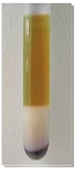
Figure 1. Clinical case. A 68-year-old man visited the emergency department of his neighborhood hospital after experiencing many weeks of exhaustion, bone pain, and night sweats. He was hospitalized due to thrombocytopenia, anemia, and hyperleukocytosis (680,000/µL). Cell settlement exhibited a pronounced buffy coat containing excessive numbers of leukemic cells. This case is highly suggestive of manifestations of leukostasis, possibly associated with extravasation and extramedullary infiltration of leukemic cells. Finally, CLL stage VI diagnosis was established.
The pathogenesis of leukostasis is still being researched, as scientists have realized the necessity of understanding the pathophysiology involved and the potential clinical implications of doing so. Increased blood viscosity is thought to play a role in the onset of leukostasis. The bulk viscosity of blood is a result of the distortion of blood cells and the bit of blood indicated by these cells at high flow rates (in larger vessels)[4]. Even with a significant rise in leukocytic cells, the overall viscosity of the blood does not appear to increase, in part due to the concomitant drop in erythrocrit with rising leukocrit. Observed leukocrits between 20% and 25% are required in cases of moderate to severe anemia with an accompanying objective increase in blood viscosity. Transfusion of erythrocytes before lowering the leukocyte count boosts the erythrocrit, which can lead to hyperviscosity. This can be exceedingly harmful to the patient, resulting in hyperviscosity clinical symptoms[5]. Erythrocytes and normal leukocytes are smaller and less deformable than leukemic blasts. When the diameter of the poorly bendable blast cell approaches that of the channel, flow in microvessels slows. Lymphoblasts are 250 to 350mm3 in size, while myeloblasts are 350 to 450mm3. It's been suggested that the larger model of myeloblasts relative to lymphoblasts answers why patients with acute myeloblastic leukemia (AML) have a higher rate of intracerebral bleeding than those with ALL. Patients with AML have leukocyte thrombi and aggregates in their cerebral vasculature, according to histopathologic studies[4]. Novotny et al.[6] and others[7] produced a score for the clinical likelihood of leukostasis. Piccirillo et al.[8] demonstrated an association between Novotny's score and early mortality in the event of a score of 3, which indicates extremely probable leukostasis. Based on their clinical symptoms, 44% to 50% of AML patients with a WBC count of >100,000/µL have a high risk of experiencing leukostasis[8]. Hyperleukocytosis is a common side effect of ALL that can occur between 10% and 30%. Rare cases of indicative leukostasis in patients with ALL often include substantially higher WBC numbers than in those with AML. Symptomatic leukostasis is extremely uncommon and only occurs in a small percentage of patients with CML or those who are experiencing a blast crisis[9]. Patients with CML frequently arrive with hyperleukocytosis and WBC counts >100,000/µL. This observation may be explained by the fact that lobulated neutrophils, metamyelocytes, and myelocytes, which are minimal and more malleable than most immature cells (blasts), are frequently seen in CML[7]. Although CLL frequently exhibits hyperleukocytosis, with total WBC counts above 100,000/µL, leukostasis-related symptoms and signs are seldom seen. Most cases of leukostasis have been observed in CLL patients with WBC levels over 1,000,000/µL[9].
The finding that there is no discernible relationship between leukocyte count and the gravity and frequency of leukostatic complications[2] suggests that leukostasis is caused by other biological processes, such as contacts between leukemic cells and the endothelium mediated by adhesion molecules[3]. In hyperleukocytosis, Bug et al.[10] and Ganzel et al.[11] reported a substantial correlation between CD11c expression and a high probability of early mortality. Furthermore, the pathophysiology of leukostasis may be influenced by cytokine-driven endothelial injury, subsequent bleeding, hypoxic damage, and leakage of AML blasts followed by further tissue destruction by matrix metalloproteases[12]. The concept of direct molecular interactions between blast cells in leukemia that lead to clumping and intravascular aggregates is being investigated as an alternative mechanism for leukostasis. The function of the molecular etiology and subsequent cell phenotype in the evolution of leukostasis is likely crucial.
3 HYPERLEUKOCYTOSIS MOLECULAR ETIOLOGY
Interestingly, different cytogenetic and molecular traits have found a correlation with promyelocytic, monocytic, and myelomonocytic AML subtypes (French-American-British classification M3/M4/M5), chromosomal (mixed lineage leukemia) MLL rearrangement 11q23, and the fms-like tyrosine kinase 3-internal tandem duplication (FLT3-ITD) mutation, all these attributes experience Hyperleukocytosis[12,13]. Cells carrying the Runt-related transcription factor 1- runt-related transcription factor 1; translocated to, 1 fusion gene have been shown to have decreased adherence to AML blasts, resulting in increased migration into the peripheral circulation[14]. Additionally, FLT3-ITD, Nucleophosmin 1, DNA (cytosine-5)-methyltransferase 3 alpha, Neuroblastoma-RAS, CCAAT/enhancer-binding protein-alpha, and tet methylcytosine dioxygenase 2 mutation frequencies were higher in 693 de novo AML patients with WBC counts greater than 50,000/µL in recent systematic review research, and hyperleukocytosis was substantially implicated to a poor prognosis[15,16]. Cytogenetically, ALL has been linked to hyperleukocytosis by the t (4;11), t (9;22), and t (1;19) mutations. There has been documented phenotypic evidence of a substantial correlation between the expression of T-cell markers and hyperleukocytosis in ALL[9].
The appearance of leukocyte-clogged blood arteries on tissue pathology is the gold standard diagnostic test for a hyperleukocytosis-related sign. Owing to the variety of this information, the diagnosis is frequently inferred and empirically treated based on knowledge of the leukocyte count, cancer type, and clinical presentation. Diagnosis imaging, such as chest plain films or computed tomography (CT) for respiratory issues and head CT or magnetic resonance imaging for neurological symptoms, should be undertaken. However, normal imaging data do not rule out hyperleukocytosis-related signs[3]. In the chemosensitive condition, the start of cytoreductive therapy must happen right away and shouldn't be postponed. Leukapheresis utilization in hyperleukocytosis and the method of cytoreduction are still up for controversy, but it is generally agreed[14].
4 CONCLUSION
Hyperleukocytosis in leukemia is correlated with boosted fatality and higher attribution of leukostasis. Furthermore, there was no correlation between age groups and hyperleukocytosis in leukemia. Although the molecular mechanisms behind leukostasis and hyperleukocytosis are still not fully understood, researchers have continued to identify the several crucial sites that play a role in regulating how leukemic blasts, genes association, and endothelial cells interact in peripheral circulation. However, further research is needed to better understand the processes through which these mutations cause hyperleukocytosis.
Acknowledgments
Not applicable.
Conflicts of Interest
The author declared no conflict of interest.
Author Contribution
Bashir BA solely contributed to the manuscript and approved the final version.
Abbreviation List
ALL, Acute lymphoblastic leukemia
AML, Acute myeloblastic leukemia
CLL, Chronic lymphocytic leukemia
CML, Chronic myeloid leukemia
DIC, Disseminated intravascular coagulation
FLT3-ITD, Fms-like tyrosine kinase 3-internal tandem duplication
TLS, Tumor lysis syndrome
WBC, White blood cell count
References
[1] Pastore F, Pastore A, Wittmann G et al. The role of therapeutic leukapheresis in hyperleukocytic AML. PLoS One, 2014; 9: e95062.[DOI]
[2] Bewersdorf JP, Zeidan AM. Hyperleukocytosis and Leukostasis in Acute Myeloid Leukemia: Can a Better Understanding of the Underlying Molecular Pathophysiology Lead to Novel Treatments? Cells, 2020; 9: 2310.[DOI]
[3] Rollig C, Ehninger G. How I treat hyperleukocytosis in acute myeloid leukemia. Blood, 2015; 125: 3246-3251.[DOI]
[4] Ali AM, Mirrakhimov AE, Abboud CN et al. Leukostasis in adult acute hyperleukocytic leukemia: A clinician’s digest. Hematol Oncol, 2016; 34: 69-78.[DOI]
[5] Giammarco S, Chiusolo P, Piccirillo N et al. Hyperleukocytosis and leukostasis: Management of a medical emergency. Expert Rev Hematol, 2016; 10: 147-54.[DOI]
[6] Novotny JR, Muller-Beissenhirtz H, Herget-Rosenthal S et al. Grading of symptoms in hyperleukocytic leukaemia: a clinical model for the role of different blast types and promyelocytes in the development of leukostasis syndrome. Eur J Haematol, 2005; 74: 501-510.[DOI]
[7] Abdullah UY, Simbak N, Azzubaidi MS et al. Hyperleucocytosis grading score and NPM1 gene mutation among patients with acute myeloid leukemia: Malaysian experience. J Hematopathol, 2020; 13: 33-40.[DOI]
[8] Piccirillo N, Laurenti L, Chiusolo P et al. Reliability of leukostasis grading score to identify patients with high-risk hyperleukocytosis. Am J Hematol, 2009; 84: 381-382.[DOI]
[9] Ali AM, Mirrakhimov AE, Abboud CN et al. Leukostasis in adult acute hyperleukocytic leukemia: a clinician’s digest. Hematol Oncol, 2016; 34: 69-78.[DOI]
[10] Bug G, Anargyrou K, Tonn T et al. Impact of leukapheresis on early death rate in adult acute myeloid leukemia presenting with hyperleukocytosis. Transfusion, 2007; 47: 1843-1850.[DOI]
[11] Ganzel C, Becker J, Mintz PD et al. Hyperleukocytosis, leukostasis and leukapheresis: Practice management. Blood Reviews, 2012; 26: 117-122.[DOI]
[12] Hatfield KJ, Reikvam H, Bruserud O. The crosstalk between the matrix metalloprotease system and the chemokine network in acute myeloid leukemia. Curr Med Chem, 2010; 17: 4448-4461.[DOI]
[13] Chaer FE, Keng M, Ballen K. MLL-Rearranged Acute Lymphoblastic Leukemia. Curr Hematol Malig Rep, 2020; 15: 83-89.[DOI]
[14] Pastore F, Pastore A, Wittmann G et al. The role of therapeutic leukapheresis in hyperleukocytotic AML. PLoS One, 2014; 9: e95062.[DOI]
[15] Saia M, Termanini A, Rizzi N et al. AML1/ETO accelerates cell migration and impairs cell-to-cell adhesion and homing of hematopoietic stem/progenitor cells. Sci Rep, 2016; 6: 34957.
[16] Tien FM, Hou HA, Tsai CH et al. Hyperleukocytosis is associated with distinct genetic alterations and is an independent poor-risk factor in de novo acute myeloid leukemia patients. Eur J Haematol, 2018; 101: 86-94.[DOI]
Copyright © 2023 The Author(s). This open-access article is licensed under a Creative Commons Attribution 4.0 International License (https://creativecommons.org/licenses/by/4.0), which permits unrestricted use, sharing, adaptation, distribution, and reproduction in any medium, provided the original work is properly cited.


