
A Case Report: Application of 68Ga-PSMA-PET/CT for the Diagnosis of Bone Metastases in Patients with Low Prostate-Specific Antigen Levels
Yilmaz Şahin1*, Ali Şahin2, Mehmet Çelik2
1Health Services Vocational School, Ataturk University, Erzurum, Turkey
2Faculty of Medicine, Department of Nuclear Medicine, Ataturk University, Erzurum, Turkey
*Correspondence to: Yilmaz Şahin, PhD, Lecturer, Therapy and Rehabilatation Department, Health Services Vocational School, Ataturk University, Erzurum 25240, Turkey; Email: yilmazsahin86@gmail.com
Abstract
Prostate cancer is the second most common type of cancer in men worldwide. It is one of the 5 most common cancer types and contributes to the greatest number of deaths among male patients. It is most common for prostate cancer to metastasize to the vertebrae, ribs, and pelvis. An important determinant of bone metastasis is the level of prostate-specific membrane antigen (PSMA). The risk of metastatic disease is low in patients who have low serum prostate-specific antigen (PSA) levels. This study examined a patient with suspected bone metastasis based on computed tomography (CT), which is considered to be a rare condition, but whose PSA level was abnormally low. 68Ga-PSMA-PET/CT scans were performed on the patient. It was discovered that bone metastases had developed in several regions of the patients’ body. Several sclerotic lesions were observed in the lumbar vertebrae and bilateral iliac wings, suggesting that bone metastases had occurred. An examination of the pelvis revealed a lesion exhibiting Ga-68 PSMA uptake, with a SUVmax of 4.97. Furthermore, laps with Ga-68 PSMA involvement with SUVmax: 8.65 could also be seen in the paracaval and aortokaval regions.
Keywords: PSMA, prostate cancer, PET/CT, bone metastasis detection
1 INTRODUCTION
Male patients are at a higher risk of developing prostate cancer than female patients. Prostate cancer ranks second among cancer-related deaths among men after lung cancer. According to studies, one in nine men will develop prostate cancer in their lifetime[1,2]. Considering the prevalence of prostate cancer cases among male patients, an early diagnosis and possible treatment are extremely important. In most cases of prostate cancer, prostate specific antigen (PSA), digital rectal examination (PMR), and multiple Transrectal Ultrasound-guided biopsies are required to diagnose the disease[3].
1.1 PSA
The PSA protein is not a cancer-specific protein, but an organ-specific antigen. As a result, it may be related to benign prostatic hyperplasia, prostatitis, and other conditions that are not malignant[4]. Conversely, prostate cancer can be detected at any PSA level. Hence, markers specific to prostate cancer are still required for the diagnosis and follow-up of the disease. As early studies revealed the presence of PSA in the serum of patients with prostate cancer, PSA began to play an increasingly important role in the diagnosis and follow-up of prostate cancer[5]. Chybowski et al. evaluated 356 patients diagnosed with prostate cancer pre-treatment since the advent of the serum PSA test, and they were among the first to evaluate the therapeutic benefits of routine bone scans[6].
1.2 Bone Metastases
Typically, men with prostate cancer will have metastases in their bones and lymph nodes. Bones provides a favorable environment for metastatic cancer cells to grow and colonize. Approximately 65% of all bone metastases originate in the breast in women and the prostate in men[7]. The advanced stages of prostate cancer can cause bone metastasis, bone pain, skeletal fractures, and even death for the patient. Detection of bone metastases is based on bone scintigraphy with a sensitivity of approximately 95%[8,9]. Computed tomography (CT) is a complementary imaging method that can be used to detect bone metastases. To detect cancer cells, Positron Emission Tomography (PET) is regarded as one of the most modern methods. By using markers such as 18F-FDG, it is possible to visualize cells with increased glucose absorption, such as metastases. PET and CT imaging can be used together to obtain high-resolution images PET and CT imaging can be used together to obtain high-resolution images[10].
1.3 68Ga-PSMA-PET/CT Imaging
As opposed to PSA, prostate-specific membrane antigen (PSMA) is a protein that is associated with glutamate carboxypeptidase II, which is an integral membrane protein. It is present in greater quantities in prostates than in other tissues (kidney, small intestine, salivary glands) and it is not released from its location[11]. 68Ga-PSMA identifies lymph nodes in patients with moderate to high-risk prostate cancer better than conventional imaging[12]. However, few literature studies have been performed on the use of 68Ga-PSMA PET/CT for evaluating bone metastases in the staging of prostate cancer.
The purpose of this study is to determine whether there are 68Ga-PSMA-PET/CT diagnostic metastases in patients with prostate cancer who have low PSA levels and no indication of bone metastases.
2 CASE REPORT
In a 92-year-old male patient undergoing a lower abdominal CT examination on January 5, 2021, multiple sclerosis was observed in an iliac wing and lumbar vertebrae with the suspicion of a bone metastasis.
An analysis of PSA was conducted on the patient on January 26, 2021, and it was observed that the PSA result was 0.00ng/mL and the fPSA result was <0.001ng/mL. Since the PSA level reference range is 0-4ng/mL, the test was repeated, but the result remained the same. In order to rule out bone metastases, it was decided to perform 68Ga-PSMA-PET/CT for the patient due to the PSA value not correlating with the suspicion of bone metastases based on CT.
Among the patient's diagnoses were prostate cancer and bone metastases. Before undergoing the 68Ga-PSMA-PET/CT scan, the patient fasted for 12h. A dose of Ga-68 PSMA of 5.29mCi was then administered intravenously to the patient. After 1h of the administration of Ga-68 PSMA, a whole-body protocol was used to obtain PET images from the skull base to the thigh level (Figure 1). The gamma-ray reduction was corrected using CT images obtained without oral contrast to determine where 68Ga-PSMA is being absorbed.
|
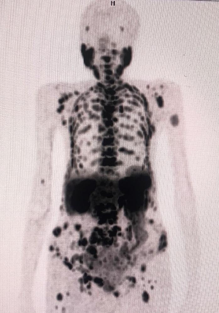
Figure 1. 68Ga-PSMA PET/CT whole body view.
3 DISCUSSION
The purpose of this study was to determine the optimal imaging tool for detecting bone metastases utilizing 68Ga-PSMA-PET/CT scans in patients with low PSA levels and no indication for bone metastases. As a result, the patient was injected with Ga-68 PSMA after PSMA was connected by in vitro method to Ga-68 obtained from the Ga-68 generator. Observations indicate that the administered radiopharmaceutical (Ga-68 PSMA) accumulates at the sites of physiological accumulation. Additionally, Lytic-sclerotic bone lesions with pathological uptake of Ga-68 PSMA were also noted in the left humerus, left clavicle, and both scapulae. A lytic-sclerotic bone lesion with pathological PSMA uptake was observed on the vertebrae and ribs, as well as numerous locations on the sacrum and pelvis (Figure 2). A lytic-sclerotic bone lesion exhibiting pathological Ga-68 PSMA involvement was observed in both proximal femurs (Figure 3). In the pelvis, a lesion showing pathological Ga-68 PSMA uptake was observed in the middle and right sections of the prostate gland, measuring approximately 34×25mm in size. Conglomerated LAPs were observed in the paracaval and aortocaval regions of the bilateral iliac chain, demonstrating pathological PSMA uptake in the perivesical, pararectal, and parasacral areas.
|

Figure 2. 68Ga-PSMA/PET-CT anterior (A) and lateral (B) view.
|
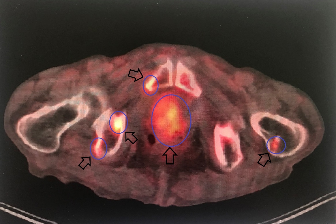
Figure 3. Prostate gland PSMA involvement.
Based on the findings of our study, 68Ga-PSMA-11 PET/CT can be a valuable imaging tool in the detection and follow-up of bone metastases in patients with metastatic prostate cancer. PET/CT imaging with 68Ga-PSMA-11 PET/CT is particularly effective for the diagnosis and assessment of patients with suspected bone metastases on CT despite low PSA levels. According to the results of this study, the 68Ga-PSMA-11 PET/CT showed a significantly higher rate of bone metastasis than the full diagnostic CT scan. These findings suggest the importance of 68Ga-PSMA-11 PET/CT for detecting bone metastases.
Acknowledgements
Not applicable.
Conflicts of Interest
There was no conflict of interest between the authors.
Author Contribution
All authors contributed to the manuscript and approved the final version.
Abbreviation List
CT, Computed Tomography
PET, Positron emission tomography
PSA, Prostate-specific antigen
PSMA, Prostate-specific membrane antigen
References
[1] Siegel RL, Miller KD, Jemal A. Cancer statistics, 2019. CA-Cancer J Clin, 2019; 69: 7-34. DOI: 10.3322/caac.21551
[2] Jemal A, Siegel R, Ward E et al. Cancer statistics, 2009. CA-Cancer J Clin, 2009; 59: 225-249. DOI: 10.3322/caac.20006
[3] Ketelsen D, Röthke M, Aschoff P et al. Detection of bone metastasis of prostate cancer-comparison of whole-body MRI and bone scintigraphy. RoFo-Fortschr Rontg Nuklearmed, 2008; 180: 746-752. DOI: 10.1055/s-2008-1027479
[4] Kosova F, Arı Z. Prostat kanseri ve apoptozis ilişkisi. J Clin Exp Invest, 2011; 2: 124-131.
[5] Papsidero LD, Wang MC, Valenzuela LA et al. A prostate antigen in sera of prostatic cancer patients. Cancer Res, 1980; 40: 2428-2432.
[6] Chybowski FM, Keller JJL, Bergstralh EJ et al. Predicting radionuclide bone scan findings in patients with newly diagnosed, untreated prostate cancer: Prostate specific antigen is superior to all other clinical parameters. J Urology, 1991; 145: 313-318. DOI: 10.1016/S0022-5347(17)38325-8
[7] Paes FM, Serafini AN. Systemic metabolic radiopharmaceutical therapy in the treatment of metastatic bone pain. Semin Nucl Med, 2010; 40: 89-104. DOI: 10.1053/j.semnuclmed.2009.10.003
[8] Hamaoka T, Madewell JE, Podoloff DA et al. Bone imaging in metastatic breast cancer. J Clin Oncol, 2004; 22: 2942-2953. DOI: 10.1200/JCO.2004.08.181
[9] Even-Sapir E, Metser U, Mishani E et al. The detection of bone metastases in patients with high-risk prostate cancer: 99mTc-MDP planar bone scintigraphy, single-and multi-field-of-view SPECT, 18F-fluoride PET, and 18F-fluoride PET/CT. J Nucl Med, 2006; 47: 287-297.
[10] Fogelman I, Cook G, Israel O et al. Positron emission tomography and bone metastases. Semin Nucl Med, 2005; 35: 135-142. DOI: 10.1053/j.semnuclmed.2004.11.005
[11] Sweat SD, Pacelli A, Murphy GP et al. Prostate-specific membrane antigen expression is greatest in prostate adenocarcinoma and lymph node metastases. Urology, 1998; 52: 637-640. DOI: 10.1016/S0090-4295(98)00278-7
[12] Lengana T, Lawal IO, Boshomane TG et al. 68Ga-PSMA PET/CT replacing bone scan in the initial staging of skeletal metastasis in prostate cancer: A fait accompli? Clin Genitourin Canc, 2018; 16: 392-401. DOI: 10.1016/j.clgc.2018.07.009
Copyright © 2022 The Author(s). This open-access article is licensed under a Creative Commons Attribution 4.0 International License (https://creativecommons.org/licenses/by/4.0), which permits unrestricted use, sharing, adaptation, distribution, and reproduction in any medium, provided the original work is properly cited.



 Copyright ©