
Increased Urinary Albumin Excretion But Less Damaged Renal Tubular Structures in Mice with Genetically Decreased Elmo1 Post Ischemia/Reperfusion Injury
Meitong Chen1, Qing Ma1, Niroshani M. W. Wariyapperuma Appuhamillage1, Yuye Wang1, Yukako Kayashima1, Nobuyo Maeda-Smithies1, Feng Li1*
1Department of Pathology and Laboratory Medicine, School of Medicine, The University of North Carolina, Chapel Hill, NC 27599, USA
*Correspondence to: Feng Li, PhD, Department of Pathology and Laboratory Medicine, The University of North Carolina, Chapel Hill, NC 27599, USA; E-mail: lif@med.unc.edu
DOI: 10.53964/id.2026004
Abstract
Renal ischemia/reperfusion injury (IRI) is a leading cause of acute kidney injury (AKI), a potentially fatal syndrome characterized by a rapid decline in kidney function. The major cause of AKI is IRI. Our prior studies have demonstrated that genetically increased Elmo1 expression in mice aggravated several kidney pathologies including diabetic nephropathy and transition of AKI to chronic kidney disease induced by IRI. However, the effects of decreased expression of Elmo1 on IRI is unclear. We compared the kidney structures and functions between wild type (WT) mice and mice with genetically decreased Elmo1 expression (Elmo1L/L) 5 days after unilateral renal IR surgery. The WT-IRI mice had typical tubular injuries including necrosis and shedding of proximal tubular cells, but these morphological changes were less severe in Elmo1L/L -IRI mice. In contrast, the urinary albumin excretion was elevated in Elmo1L/L -IRI mice compared with WT counterparts. While the expression of inflammatory markers (e.g., Il6, Tnfa, Cxcl1 and Tlr4) was comparable between WT and Elmo1L/L mice with IRI which significantly higher than control mice, the expression of antioxidant markers (e.g., Sod1 and Sod2) was preserved in Elmo1L/L -IRI mice which was significantly decreased in WT-IRI mice. We conclude that Elmo1L/L mice had preserved tubular structures but increased urinary albumin excretion after IRI, suggesting that the role of Elmo1 in IRI is complex and it merits future evaluation.
Keywords: low Elmo1 expression, mice, ischemia reperfusion injury
1 INTRODUCTION
Acute kidney injury (AKI) is a clinical condition associated with high morbidity and mortality[1,2]. One of the leading causes of AKI is kidney ischemia/reperfusion injury (IRI), which occurs when the blood supply to the kidney is temporarily blocked then restored[3,4] . While reperfusion is essential to re-establish oxygen delivery, the sudden reintroduction of oxygen reacts with accumulated metabolic intermediates from the ischemic phase, causing a rapid increase in superoxide and other reactive oxygen species (ROS)[5,6]. This initial oxidative burst is further amplified by impaired mitochondrial electron transport and activation of NADPH oxidases (Nox), which sustains ROS generation during the injury and repair processes[7,8]. In AKI, excessive ROS damages renal proximal tubular cells mainly and contributes to both acute and chronic renal injury[9,10].
Engulfment and cell motility protein 1 (Elmo1) was first discovered in Caenorhabditis elegans as the CED-12 protein for its role on cell corpse engulfment which helps internalize apoptotic cells. It was first identified as part of the CED-2/CED-5/CED-12 signaling pathway in which cytoskeletal remodeling occurred during phagocytosis and cell migration[11]. Later, researchers found that it was a cytoplasmic adaptor protein involved in regulating cytoskeletal dynamics, phagocytosis, and cell survival signaling. Genome-wide association studies have identified multiple single-nucleotide polymorphisms in the ELMO1 gene that are strongly associated with kidney disease including diabetic nephropathy[12,13] and nephrotic syndrome[14]. Elmo1 has also been implicated in modulating oxidative stress pathways. Previous work has demonstrated that increase in Elmo1 expression (200% of normal) linked to increased ROS generation, which aggravated diabetic complications, while decreased Elmo1 expression (30%) had protective effects in diabetic mice[15,16].
Our previous studies also showed that Elmo1 overexpression exacerbated the severity of IRI-induced kidney disease[17]. However, the effects of reduced Elmo1 expression on IRI is unknown. Because (1) Elmo1 regulates autophagy induction[18] and excessive or insufficient autophagy is observed during kidney disease progression[19,20], (2) Elmo1 deficiency causes cell cycle arrest and deceased proliferation[21] and cell proliferation is critical for kidney repair after IRI[3], it is important to investigate the role of Elmo1 deficiency in IRI.
In the current study, we applied renal unilateral IR surgery to mice with decreased expression of Elmo1 and found that haploinsufficiency for Elmo1 led to increased urinary albumin excretion but less damaged tubular structures in acute phase of IRI (5 days after surgery) in mice.
2 MATERIALS AND METHODS
2.1 Mice
Male littermates of WT and Elmo1L/L (C57BL/6J background) at ages 12-15 weeks were used[15]. Mice were maintained under standard housing conditions with a 12-hour light/dark cycle and free access to food and water. All animal procedures complied with the National Institutes of Health guidelines for the care and use of laboratory animals and were approved by the Institutional Animal Care and Use Committee at the University of North Carolina at Chapel Hill (Protocol #: 25-190). WT or Elmo1L/L Mice were randomly enrolled into different groups.
2.2 Renal IRI Procedure
The surgery was performed on mice using a dorsal approach. Mice were anesthetized with 1.5% isoflurane inhalation, and body temperature was maintained at 37 °C throughout the procedure. Following anesthesia, the surgical site was disinfected, a small incision was made through the muscle fascia to expose the left kidney. The left renal pedicle was occluded using a microaneurysm clamp for 30 minutes to induce ischemia (successful ischemia was confirmed by the kidney turning dark). After clamp removal, reperfusion was initiated and the incision was closed. The muscle fascia was closed with two interrupted absorbable sutures, and the skin was closed using wound clips[22]. Intact WT and Elmo1L/L mice were included as respective controls.
2.3 Biochemical Analysis
Spot urines were collected 5 days after surgery, prior to euthanasia. Following euthanasia, blood and kidneys were collected. Plasma cystatin C and urinary albumin were measured by ELISA kits (MSCTC0, R&D system; Albuwell M kit #1011, Ethos Biosciences). Mouse urine creatinine was measured by an enzymatic assay kit (80350, Crystal Chem)[22]. The experiments were done by investigators who were blinded to the experimental groups
2.4 Histological Examination
Kidney tissues (both affected and contralateral) were fixed in 4% paraformaldehyde, embedded in paraffin, sectioned (5µm), and stained using the Periodic acid–Schiff (PAS). Images were captured with cellSENS Microscope Imaging Software (version 4.3, Evident Olympus)[17]. Semi-quantification of tubular injury was scored using a scale of 0 to 4: 0, no injury; 1. <25%; 2. 25-50%; 3. 50-75%; 4. >75%. Scoring was done by an investigator who was blinded to the experimental groups[22].
2.5 Quantitative RT-PCR
Total RNA from tissues was extracted using Trizol (Life Technologies) following the manufacturer's instructions. NanoDrop spectrophotometer method and gel electrophoresis were used to check quantity and quality of RNA. mRNA was quantified with QuantStudio 3 Real-Time PCR system (Thermo Fisher Scientific) by using one-step RT-PCR Kit (Bio Rad) with Actb as reference gene[22]. All used qRT-PCR primers and probes were shown in Table 1. The experiments were conducted by investigators who were blinded to the experimental groups.
Table 1. qRT-PCR Primers and Probes List
Gene |
|
Sequence (5’-3’) |
Cxcl1 |
Forward |
GCT TGT AGG TGT TGC CCT C |
|
Reverse |
AGG CAA GCC TCG CGA CCA T |
|
Probe |
FAM-AC CCA AAC CGA AGT CAT AGC CAC ACT C— TAMRA |
Cubn |
Forward |
TTT GGA CCG TTC TGT GGC AT |
|
Reverse |
GGA ATC TTA TGA AGA CCC GA |
|
Probe |
FAM-AC ACT GTG GTA GCA CCC TTT CAT GC— TAMRA |
Edn1 |
Forward |
CAG CAG TTA GTG AGA GGA AG |
|
Reverse |
GAC GCT GTT TCT CAT GGT CT |
|
Probe |
FMA-TC CCG AGC GCG TCG TAC CGT ATG- TAMRA |
Il6 |
Forward |
CTC TCT GCA AGA GAC TTC CA |
|
Reverse |
CTC TCC GGA CTT GTG AAG TA |
|
Probe |
FAM-CTG ATG CTG GTG ACA ACC ACG GCC T—TAMRA |
Lrp2 |
Forward |
GAA TCC GCC AAG ACA CTG AT |
|
Reverse |
ACA AGA GTC CCC TGG GCA TA |
|
Probe |
FAM-CCCTCG CTC AGC TGC TTC CCG TA— TAMRA |
Nox2 |
Forward |
TGCCACCAGTCTGAAACTCA |
|
Reverse |
CAGCAGGTCTGCAAACCACT |
|
Probe |
FAM-AGGCATGCGTGTCCCTGCACAGCCA-TAMRA |
Nphs1 |
Forward |
CAG CTG CTA GTC TGC GAG G |
|
Reverse |
ATC AAT GAC AGG AGG TCC TG |
|
Probe |
FAM-TC CAA CCC AGC CTT GGC CAC TC- TAMRA |
Sod1 |
Forward |
CCA TTG AAG ATC GTG TGA TCT C |
|
Reverse |
CTT GTT TCT CAT GGA CCA CC |
|
Probe |
FAM-CA GGA GAG CAT TCC ATC ATT GGC CG—TAMRA |
Sod2 |
Forward |
AAG GAA CAA GGT CGC TTA CA |
|
Reverse |
AGC AGC GGA ATA AGG CCT GT |
|
Probe |
FAM-TG CTG CCT GCT CTA ATC AGG ACC CA—TAMRA |
Sod3 |
Forward |
CCT TCT TGT TCT ACG GCT TG |
|
Reverse |
CTG GAC TCC CCT GGA TTT GA |
|
Probe |
FAM-TG ACA GAG CCA CAG GCC GCC AGT—TAMRA |
Tlr4 |
Forward |
GGT GAG AAA TGA GCT GGT AAA G |
|
Reverse |
GCA ATG GCT ACA CCA GGA AT |
|
Probe |
FAM-TG CCC CGC TTT CAC CTC TGC CTT CA—TAMRA |
Tnfα |
Forward |
CAC ACT CAG ATC ATC TTC TCA A |
|
Reverse |
AGC TGC TCC TCC ACT TGG T |
|
Probe |
FAM-AG CCT GTA GCC CAC GTC GTA GCA--TAMRA |
Wt1 |
Forward |
CCA GTG AGA AAC GTC CTT TC |
|
Reverse |
GGT CAC TCT TTG CAG GAA AG |
|
Probe |
FAM-CACGCCGCACATAGACCTCACC TAMRA |
Actb |
Forward |
AAG AGC TAT GAG CTG CCT GA |
|
Reverse |
TGA TGG AAT TGA ATG TAG TTT CA |
|
Probe |
TET-CAC TAT TGG CAA CGA GCG GTT CCG-TAMRA |
2.6 Statistical Analysis
The sample size was determined by power calculation based on our previous study[17,22]. Data are shown as mean±SEM. The two-way ANOVA was performed using JMP version 17.2.0 (SAS Institute Inc.). Two-tailed Tukey–Kramer HSD test was used for post hoc comparison. All Statistical significance was set at P<0.05.
3.1 The Kidney Tubular Structure Was Improved in Elmo1L/L Mice 5 Days Post IRI
IRI causes structural and functional losses in acute and chronic kidney injury[17,22] . To evaluate how reduced Elmo1 expression influences the structures of kidneys after IRI acutely, we first examined the renal tubular structures of affected kidneys. As expected, WT-IRI mice showed protein casts and shedding of proximal tubular cells[22] . Surprisingly, Elmo1L/L-IRI showed only minimal renal injury compared with WT counterparts. The kidney structures were not different between WT-control and ElmoL/L -control mice (Figure 1).
|
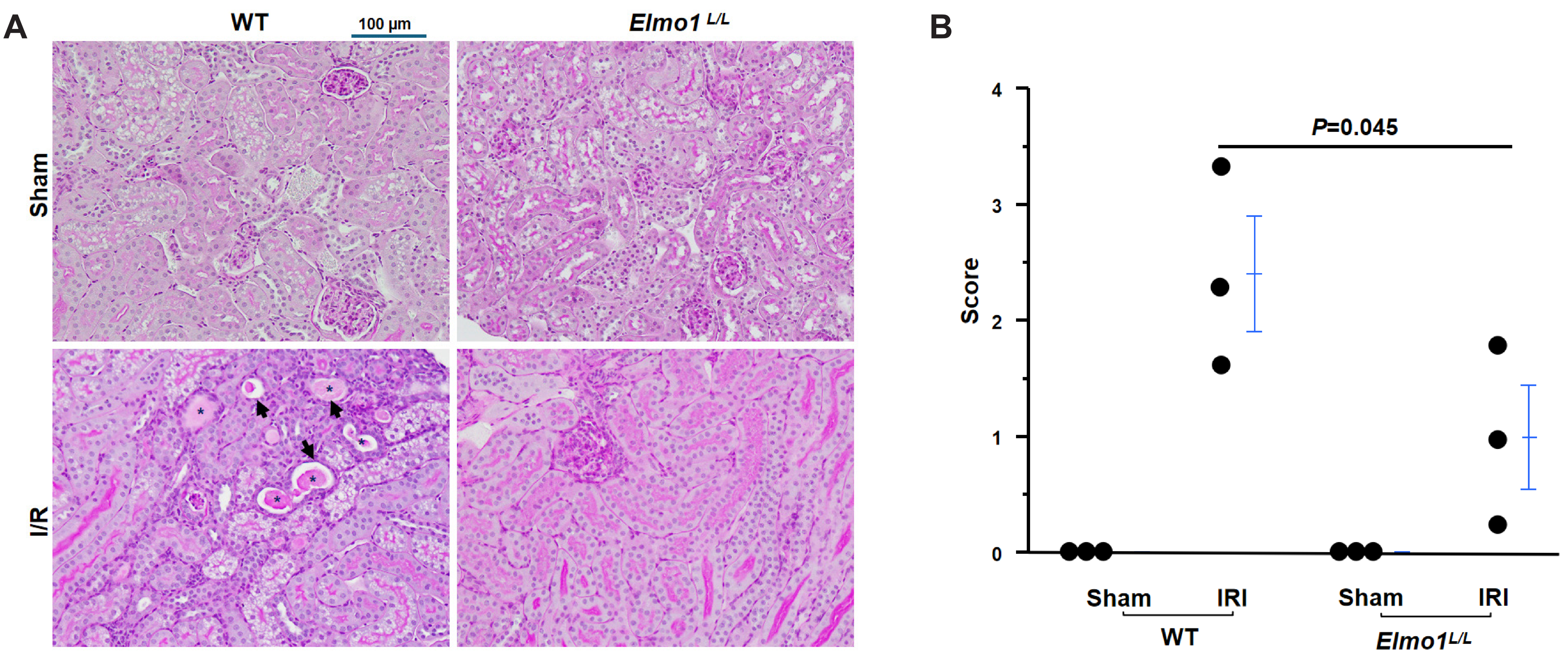
Figure 1. The Tubular Structure is Less Damaged in Elmo1L/L Mice than WT Mice 5 Days Post IRI. A: Representative periodic acid–Schiff (PAS) stain of the affected kidneys in the four groups of mice. Asterisk: protein cast. Arrow: necrotic tubules. B: Semi-quantitative injury score of the affected kidneys of the four groups of mice. C. Control. n=3.
The structures of the glomeruli from mice with IRI did not show obvious abnormalities examined by a light microscope (Figure 2A). The expression of markers of podocytes [Wt1 encoding Wilms' Tumor 1) and Nphs1(encoding Nephrin) was determined by qRT-PCR. While the expression of Wt1 was not different between four groups of mice (Figure 2B), the expression of Nphs1 tended to decrease in both WT and ElmoL/L mice with IRI compared with control mice and there was no difference between WT-IRI and ElmoL/L-IRI mice (Figure 2C).
|
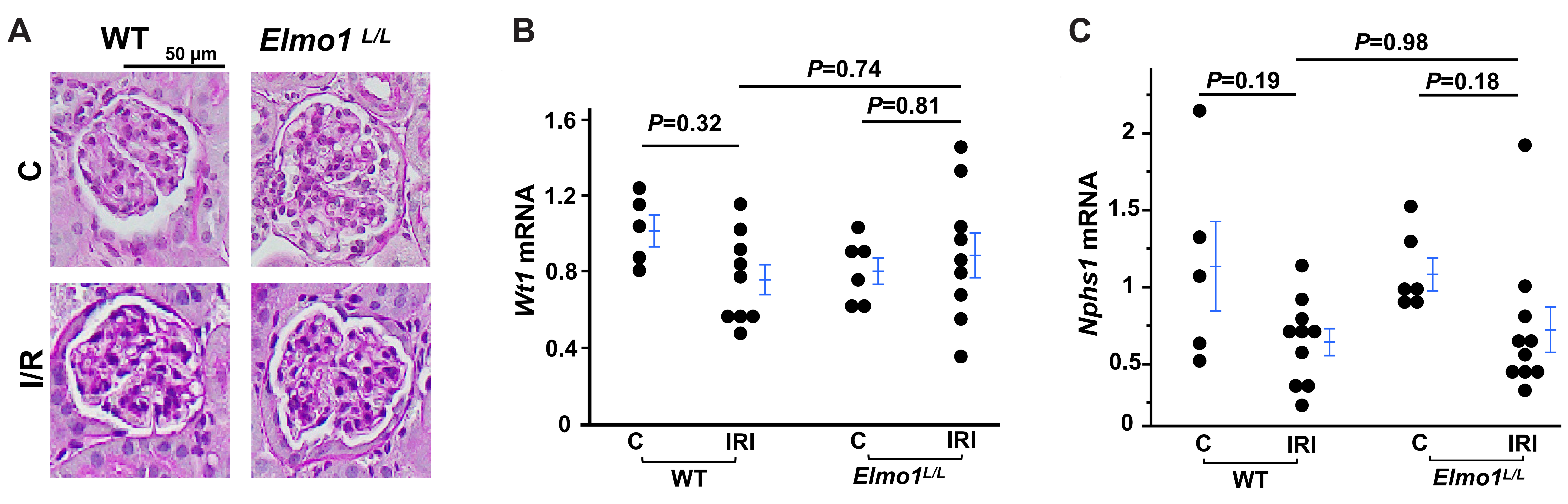
Figure 2. There are No Obvious Abnormalities in Glomeruli 5 Days Post IRI. A: Representative periodic acid–Schiff (PAS) stain of glomerulus in the four groups of mice. B-C: The expression markers of podocytes. Wt1: Encoding Wilms' tumor 1, Nphs1: Encoding nephrin. C. Control. n=5-10.
The right kidney weight (unaffected)/body weight ratio was lower in ElmoL/L mice with IRI compared with WT counterparts (Table 2), however, the structures of contralateral kidneys examined by a light microscope did not reveal obvious difference between four groups of mice (Figure 3).
Table 2. General Characteristics of Four Groups of Mice
Characteristics |
WT (control) |
Elmo1L/L (control) |
WT (IRI) |
Elmo1L/L (IRI) |
n |
5 |
6 |
10 |
10 |
Body weight (g) |
27.71 0.92 |
25.44 1.55 |
26.82 0.50 |
23.80 0.99 |
Left kidney (affected) (mg) |
155.2 4.69 |
143.17 5.51 |
176.1 5.74 |
142.7 8.56 |
Right kidney (mg) |
158.8 2.74 |
149.67 9.39 |
205.3 6.83 |
151.4 11.17 |
Heart (mg) |
115.2 2.25 |
118.67 4.13 |
131.4 3.82 |
116.6 5.33 |
Liver (g) |
1.22 0.04 |
1.16 0.04 |
1.37 0.03 |
1.23 0.05 |
Left kidney/BW (%) |
0.56 0.016 |
0.57 0.031 |
0.66 0.019 |
0.60 0.017 |
Right kidney/BW (%) |
0.58 0.19 |
0.59 0.017 |
0.77 0.027* |
0.63 0.027 |
Heart/BW (%) |
0.42 0.018 |
0.47 0.028 |
0.49 0.014 |
0.49 0.07 |
Liver/BW (%) |
4.44 0.32 |
4.60 1.53 |
5.10 1.08 |
5.15 0.58 |
Notes: Body weight (BW) and organ weight of four groups of mice were measured on day 5 after surgery. *P<0.05 vs. other groups.
|
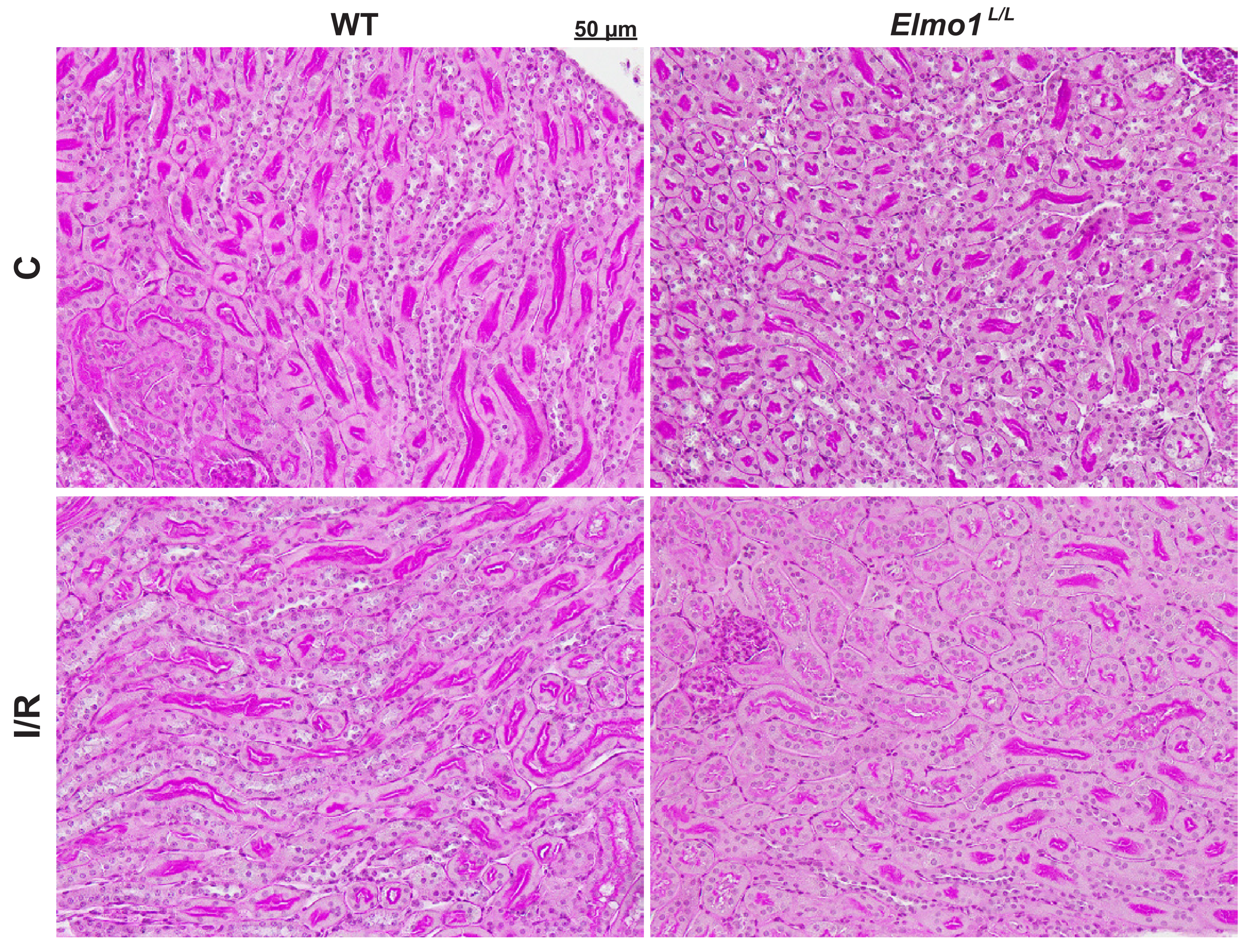
Figure 3. Ther is No Obvious Abnormality of the Tubular Structure in the contralateral kidneys from WT and Elmo1L/L Mice 5 Days Post IRI. Representative PAS stain of kidneys in the four groups of mice. C. Control.
3.2 Elmo1L/L Mice with IRI Had Higher Urinary Albumin Excretion than WT Counterparts 5 Days Post IRI
Since IRI caused a decline in kidney function[22], we measured two markers of kidney function: plasma cystatin C and urinary albumin-to-creatinine ratio (UACR)[23-25] . In WT mice, plasma cystatin C slightly increased after IRI compared with WT control mice (Figure 4A). Elmo1L/L -IRI mice had a significant increase in cystatin C compared with Elmo1L/L control mice, and higher than WT-IRI mice as well. WT-IRI mice had an increase in UACR compared with WT control mice as expected. Elmo1L/L -IRI mice had higher UACR compared with WT counterparts (Figure 4B). The levels of cystatin C and UACR were not different between two groups of control mice (Figure 4).
|
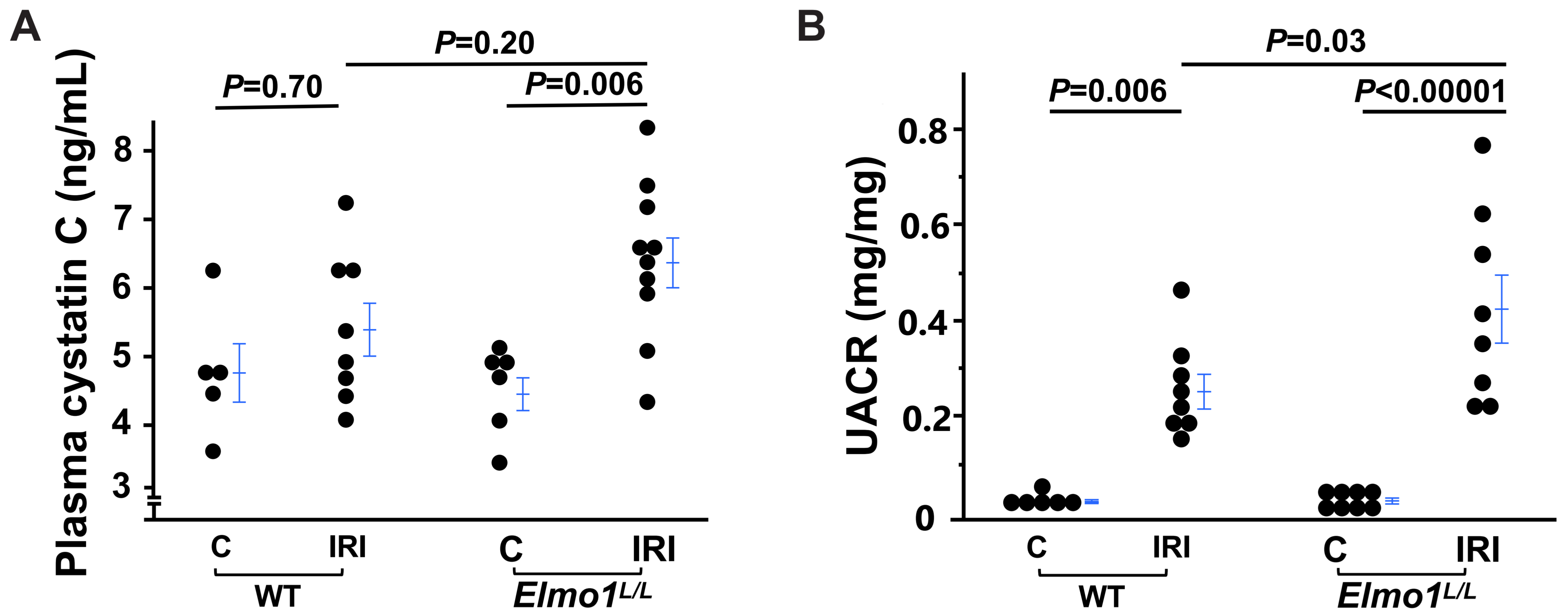
Figure 4. The Declined Kidney Function by IRI is Aggravated in Elmo1L/L Mice 5 Days Post IR Surgery. A: The plasma cystatin C concentration. B: Urinary albumin excretion (albumin/creatinine ratio, UACR). C.Control. n=5-10.
3.3 The expression of inflammatory markers was not different between WT-IRI and Elmo1L/L-IRI mice
Inflammation response plays pivotal roles in the IRI process[26,27]. To determine whether low Elmo1 alters inflammatory responses to IRI, we evaluated the mRNA levels of Il6, Tnfα, Cxcl1 and Tlr4 in the affected kidneys. Compared with their respective control groups, both WT and Elmo1L/L mice showed significant increases in the mRNA levels of these markers of inflammation following IRI. There was no difference between WT and Elmo1L/L mice with IRI (Figure 5).
|
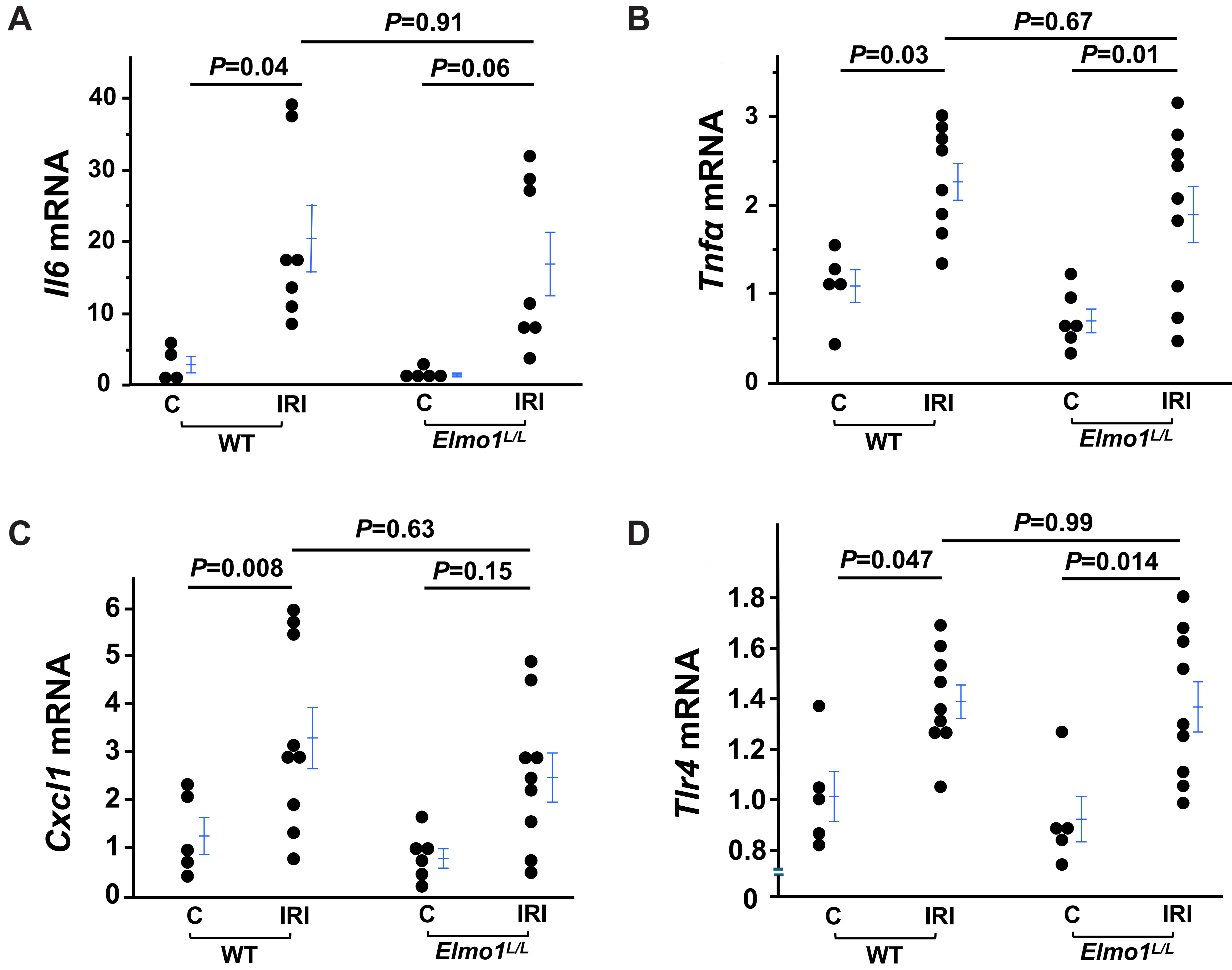
Figure 5. The Expression of Inflammatory Markers Examined in Affected Kidneys is Not Different between WT and Elmo1L/L Mice 5 Days Post IRI. C.Control. n=5-10.
3.4 The Expression of Antioxidant Enzymes was Preserved in Elmo1L/L -IRI Mice
Because prior studies show antioxidant enzymes decreased in kidneys with IRI[22,28], we examined the expression of antioxidant enzymes: superoxide dismutase 1/2/3 (Sod1, Sod2 and Sod3) in affected kidneys. In WT mice, the expression of Sod1 , Sod2 and Sod3 was reduced after IRI as expected. However, this decrease of Sod1 and Sod2 was not observed in Elmo1L/L mice with IRI, only the expression of Sod3 (extracellular SOD) was decreased in Elmo1L/L mice with IRI. Rac1-GTP dependent NADPH oxidase (Nox2) mRNA level was increased in WT mice with IRI consistent with previous reporting[5,29]. Interestingly, the expression of Nox2 increased in Elmo1L/L mice with IRI as well, and there was no difference between WT and Elmo1L/L mice with IRI (Figure 6).
|
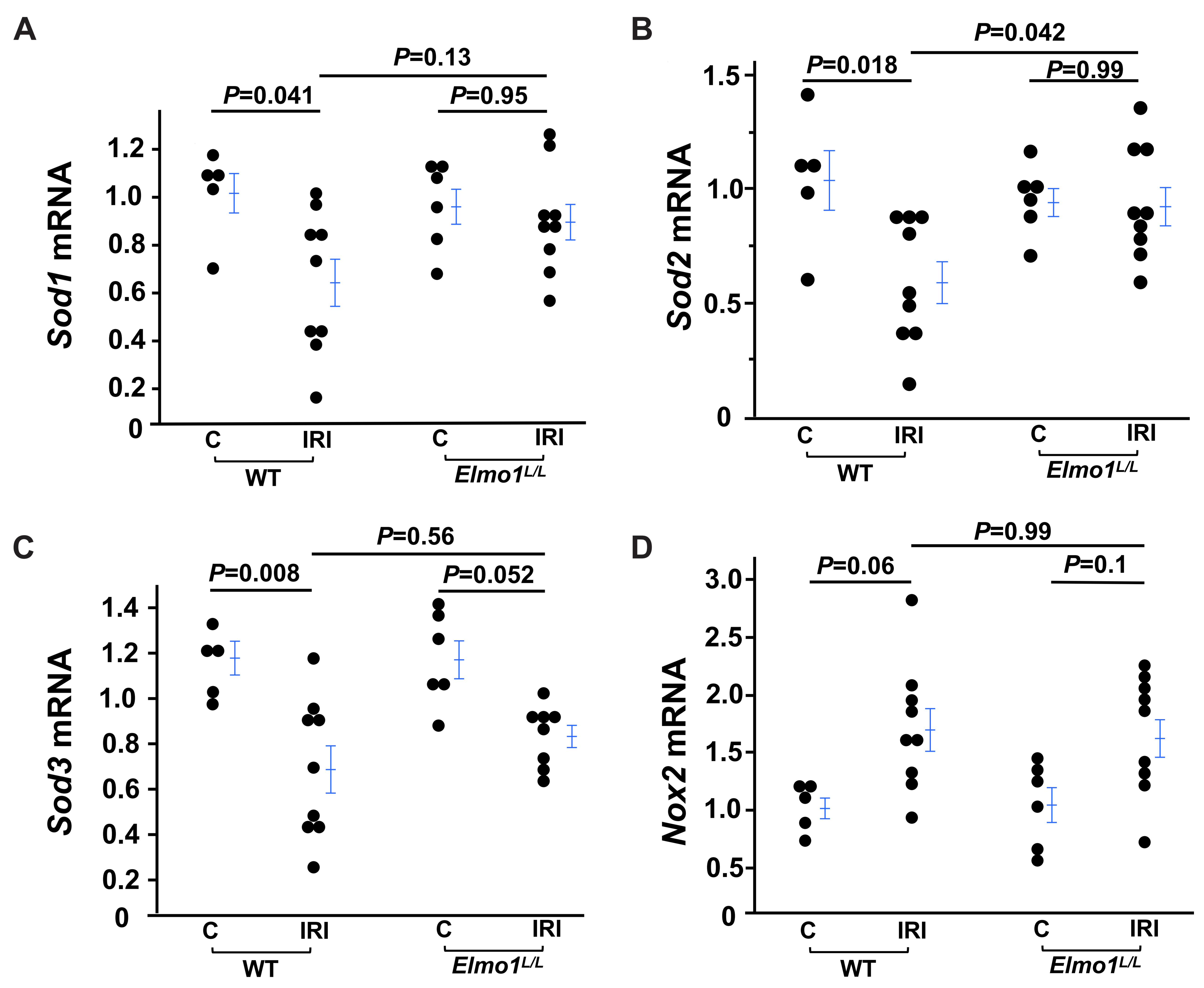
Figure 6. The Expression of Sod1 and Sod2 is Preserved in Affected Kidneys from Elmo1L/L Mice 5 Days Post IRI. mRNA levels of Sod1 (A), Sod2 (B), Sod3 (C) and Nox2 (D). C. Control. n=5-10.
Because proximal tubule protein uptake is mediated by 2 receptors, megalin and cubulin[30,31], we determined the expression of Lrp2 (encoding megalin) and Cubn (encoding cubulin) and found the expression of them was not different in affected kidneys from four groups of mice (Figure 7),
|
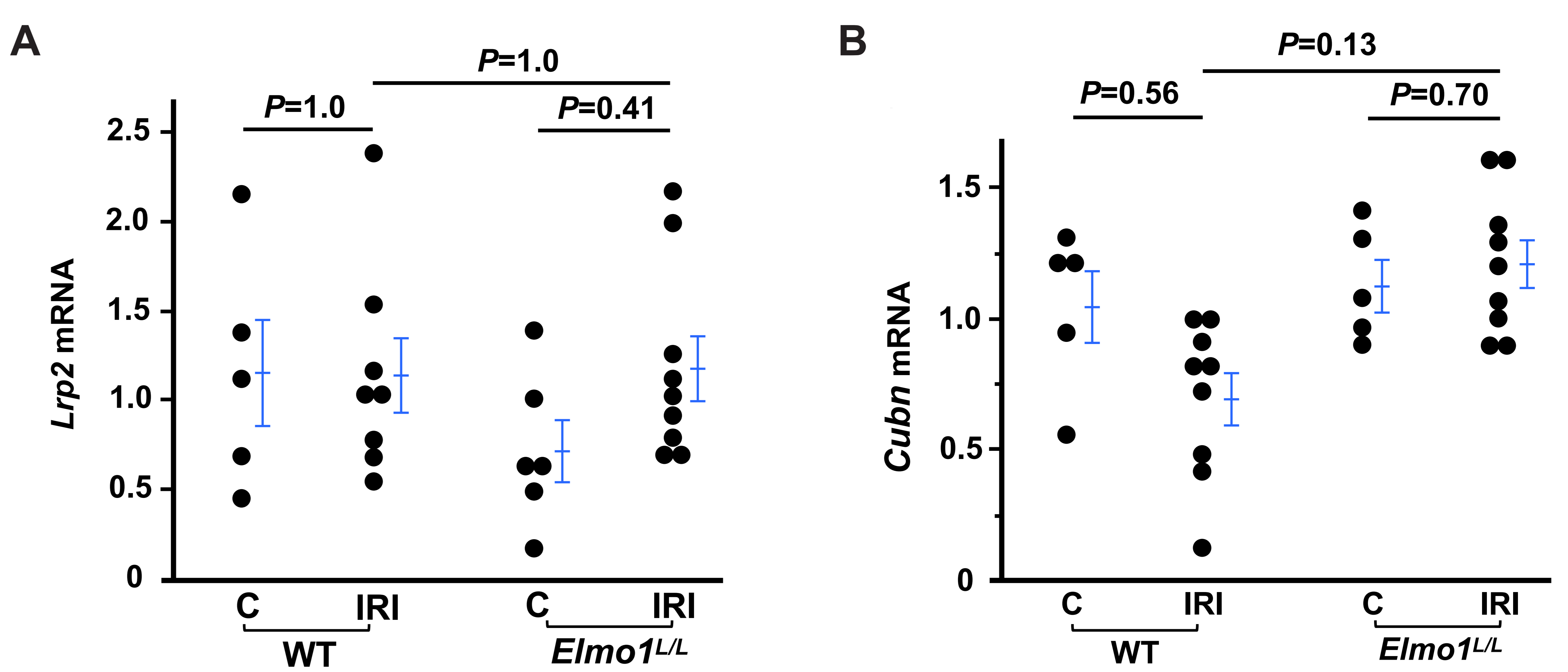
Figure 7. The Expression of Lrp2 (Encoding Megalin) and Cubn (Encoding Cubulin) in Affected Kidneys from Four Groups of Mice. C. Control. n=5-10.
3.5 The expression of Edn1 was not different between WT-IRI and Elmo1L/L -IRI mice
Endothelin-1 (ET-1) is involved in ischemic renal damage[32,33], and Elom1 is associated with Edn1 (encoding pre-pro-ET-1) expression (GSE299038[21]). Therefore, we determined the expression of Edn1 in the affected kidneys and found that the expression of Edn1 increased in both WT and Elmo1L/L mice with IRI compared with their respective controls, and there was no difference between WT and Elmo1L/L mice with IRI (Figure 8).
|
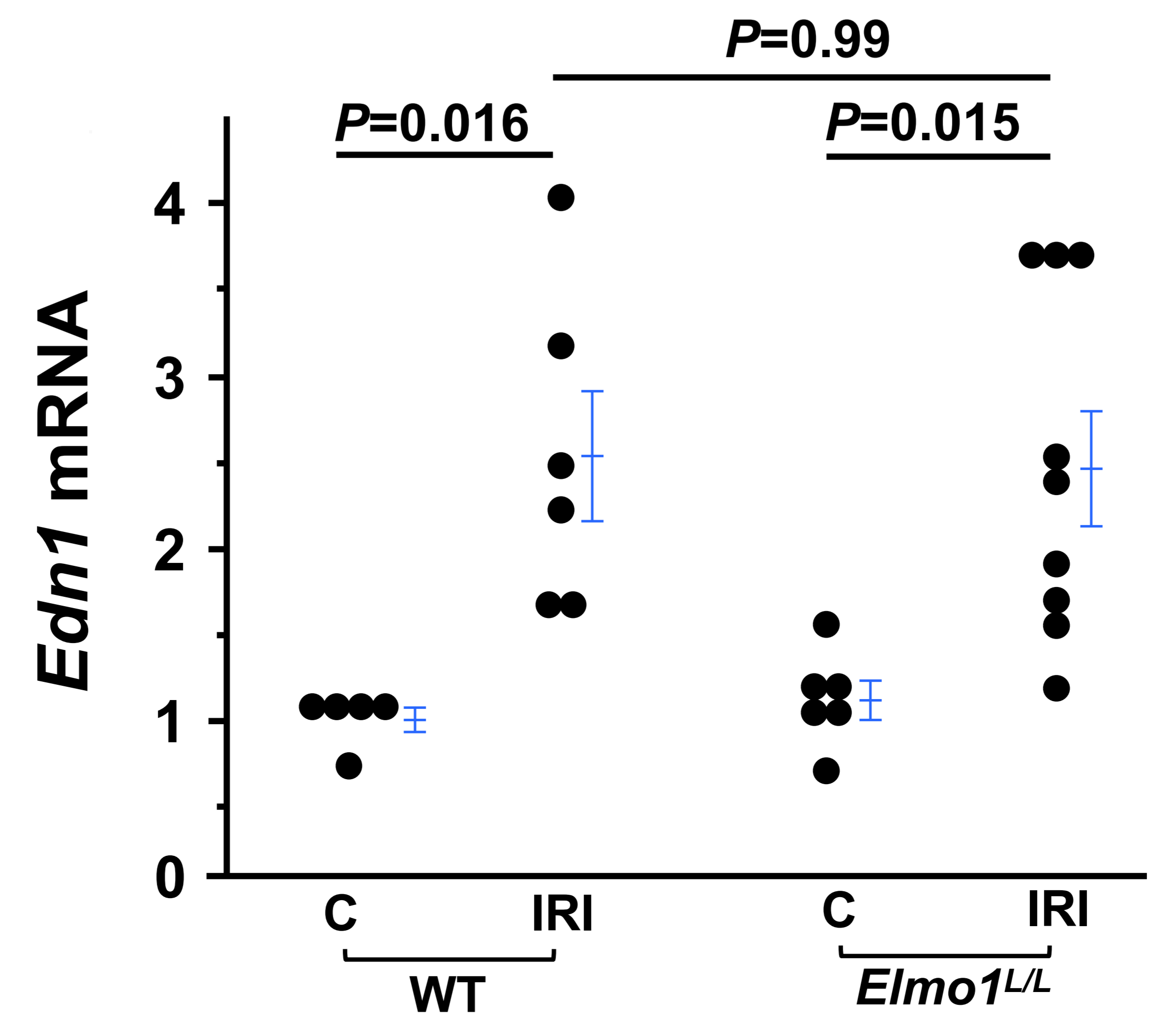
Figure 8. The Expression of Edn1 in the Affected Kidneys from Elmo1L/ L is Not Higher that WT Counterparts 5 Days Post IRI. C.Control. n=5-9.
4 DISCUSSION
In this study, we characterized the effects of genetically reduced Elmo1 expression on renal outcomes following IRI, assessing structural injury, functional impairment, and the expression of markers of inflammation and antioxidant. Improved tubular structure was observed in Elmo1L/L mice following IRI. The kidney function of Elmo1L/L -IRI mice was worse compared with WT counterparts, as evidenced by higher urinary albumin excretion. The expression of markers of inflammation (e.g., Il6 and Tnfα) was increased similarly in WT and Elmo1L/L mice with IRI. The expression of antioxidants (e.g., Sod1 and Sod2) decreased in WT-IRI mice but not in Elmo1L/L -IRI mice.
Prior studies demonstrated that antioxidant enzymes Sod1 and Sod2 are reduced in the kidneys of WT mice following IRI[22] . We confirmed this in our study. In contrast, Elmo1L/L-IRI mice had the same levels of Sod1 and Sod2 as control groups. The expression of inflammatory markers was not different between WT and Elmo1L/L mice with IRI, which significantly increased compared with control mice. Taken together, the preserved antioxidative response could contribute to the less tubular injury observed in Elmo1L/L-IRI mice.
A number of important limitations of the present study need to be considered. The morphological examination was performed by using a light microscope, which can not detect the changes of ultrastructures of glomeruli and tubules, especially podocytes. Further studies using EM are warranted to elucidate the alteration of ultrastructures of kidneys with IRI. Although we did not find obvious abnormality of the tubular structures of contralateral kidneys in WT and Elmo1L/L mice with IRI, the functional analysis of these kidneys is needed. We acknowledge as a limitation of the study that we only measured the mRNA levels of megalin and cucblin.
5 CONCLUSION
In summary, mice with decreased expression of Elmo1 had less tubular injury but more severe kidney dysfunction 5 days post IRI. The data prompt us to hypothesize that low Elmo1 may have different effects on different types of cells in the kidneys. Future comprehensive stereological and/or single cell RNA-seq analysis may provide insight into the complex effects of Elmo1 on AKI.
Acknowledgements
This work was supported by grants from the National Institutes of Health (R01HL049277 to N.M-S., R01HD101485 to F.L).
Conflicts of Interest
The authors declared no conflict of interest.
Ethical Statement
All animal procedures were approved by the Institutional Animal Care and Use Committee (IACUC) at the University of North Carolina at Chapel Hill (Protocol #: 25-190).
Copyright Permissions
Copyright © 2026 The Author(s). Published by Innovation Forever Publishing Group Limited. This open-access article is licensed under a Creative Commons Attribution 4.0 International License (https://creativecommons.org/licenses/by/4.0), which permits unrestricted use, sharing, adaptation, distribution, and reproduction in any medium, provided the original work is properly cited.
Data Availability
All data generated or analyzed during this study are included in this published article.
Author Contribution
Li F designed the study. Li F, Chen M, Ma Q, Wariyapperuma Appuhamillage NMW, Wang Y, and Kayashima Y carried out the experiments. Li F, Ma Q, Wariyapperuma Appuhamillage NMW, Chen M, and Maeda-Smithies N analyzed and interpreted the data. Li F and Chen M drafted the paper, and Li F, Kayashima Y, and Maeda-Smithies N revised it. All authors have read and agreed to the published version of the manuscript.
Abbreviation List
AKI, Acute kidney injury
BW, Body weight
Elmo1, Engulfment and cell motility protein 1
EM, Electron microscope
ET-1, Endothelin-1
IRI, Ischemia/reperfusion injury
Nox, NADPH oxidases
PAS, Periodic acid–Schiff
ROS, Reactive oxygen species
UACR, Urinary albumin-to-creatinine ratio
WT, Wild type
References
[1] Turgut F, Awad AS, Abdel-Rahman EM. Acute Kidney Injury: Medical Causes and Pathogenesis. J Clin Med, 2023; 12: 375.[DOI]
[2] Hobson CE, Yavas S, Segal MS et al. Acute kidney injury is associated with increased long-term mortality after cardiothoracic surgery. Circulation, 2009; 119: 2444-2453.[DOI]
[3] Bonventre JV, Yang L. Cellular pathophysiology of ischemic acute kidney injury. J Clin Invest, 2011; 121: 4210-4221.[DOI]
[4] Malek M, Nematbakhsh M. Renal ischemia/reperfusion injury; from pathophysiology to treatment. J Renal Inj Prev, 2015; 4: 20-27.[DOI]
[5] Granger DN, Kvietys PR. Reperfusion injury and reactive oxygen species: The evolution of a concept. Redox Biol, 2015; 6: 524-551.[DOI]
[6] Tejchman K, Kotfis K, Sienko J. Biomarkers and Mechanisms of Oxidative Stress-Last 20 Years of Research with an Emphasis on Kidney Damage and Renal Transplantation. Int J Mol Sci, 2021; 22: 8010.[DOI]
[7] Dikalov S. Cross talk between mitochondria and NADPH oxidases. Free Radic Biol Med, 2011; 51: 1289-1301.[DOI]
[8] Bedard K, Krause KH. The NOX family of ROS-generating NADPH oxidases: physiology and pathophysiology. Physiol Rev, 2007; 87: 245-313.[DOI]
[9] Basile DP, Anderson MD, Sutton TA. Pathophysiology of acute kidney injury. Compr Physiol, 2012; 2: 1303-1353.[DOI]
[10] Chevalier RL. The proximal tubule is the primary target of injury and progression of kidney disease: role of the glomerulotubular junction. Am J Physiol Renal Physiol, 2016; 311: F145-F161.[DOI]
[11] Gumienny TL, Brugnera E, Tosello-Trampont AC et al. CED-12/ELMO, a novel member of the CrkII/Dock180/Rac pathway, is required for phagocytosis and cell migration. Cell, 2001; 107: 27-41.[DOI]
[12] Shimazaki A, Kawamura Y, Kanazawa A et al. Genetic variations in the gene encoding ELMO1 are associated with susceptibility to diabetic nephropathy. Diabetes, 2005; 54: 1171-1178.[DOI]
[13] Hanson RL, Millis MP, Young NJ et al. ELMO1 variants and susceptibility to diabetic nephropathy in American Indians. Mol Genet Metab, 2010; 101: 383-390.[DOI]
[14] Hassan EA, Elsaid AM, Abou-Elzahab MM et al. The Potential Impact of MYH9 (rs3752462) and ELMO1 (rs741301) Genetic Variants on the Risk of Nephrotic Syndrome Incidence. Biochem Genet, 2024; 62: 1304-1324.[DOI]
[15] Hathaway CK, Chang AS, Grant R et al. High Elmo1 expression aggravates and low Elmo1 expression prevents diabetic nephropathy. Proc Natl Acad Sci USA, 2016; 113: 2218-2222.[DOI]
[16] Kakoki M, Bahnson EM, Hagaman JR et al. Engulfment and cell motility protein 1 potentiates diabetic cardiomyopathy via Rac-dependent and Rac-independent ROS production. JCI Insight, 2019; 4: e127660.[DOI]
[17] Zhou J, Wang Y, Hagaman J et al. Vitamin B12 Protects the Exacerbated Ischemia-Reperfusion Injury-Induced Chronic Kidney Disease in Mice with Genetically Increased Elmo1. Antioxidants -Basel, 2025; 14: 1277.[DOI]
[18] Sarkar A, Tindle C, Pranadinata RF et al. ELMO1 Regulates Autophagy Induction and Bacterial Clearance During Enteric Infection. J Infect Dis, 2017; 216: 1655-1666.[DOI]
[19] Li Y, Ma T, Liang X et al. Non-coding RNAs regulate autophagy in kidney disease: friend or foe? Autophagy, 2025; 21: 2537-2560.[DOI]
[20] Zhou G, Zhang L, Bai W et al. Devil or angel: A review of autophagy in ischemia-reperfusion injury. Medicine, 2025; 104: e45330.[DOI]
[21] Kayashima Y, Deshmukh AA, Kiyokawa Y et al. Downregulation of Engulfment and cell motility 1 (Elmo1) induces quiescence and resistance to poly(I:C)-induced apoptosis in endothelial cells. Cell Death Dis, 2025; 16: 262.[DOI]
[22] Li F, Bahnson EM, Wilder J et al. Oral high dose vitamin B12 decreases renal superoxide and post-ischemia/reperfusion injury in mice. Redox Biol, 2020; 32: 101504.[DOI]
[23] Gowda S, Desai PB, Kulkarni SS et al. Markers of renal function tests. N Am J Med Sci, 2010; 2: 170-173.
[24] West M, Kirby A, Stewart RA et al. Circulating Cystatin C Is an Independent Risk Marker for Cardiovascular Outcomes, Development of Renal Impairment, and Long-Term Mortality in Patients With Stable Coronary Heart Disease: The LIPID Study. J Am Heart Assoc, 2022; 11: e020745.[DOI]
[25] de Zeeuw D, Hillege HL, de Jong PE. The kidney, a cardiovascular risk marker, and a new target for therapy. Kidney Int Suppl, 2005; 68: S25-S29.[DOI]
[26] Lu CY, Winterberg PD, Chen J et al. Acute kidney injury: a conspiracy of Toll-like receptor 4 on endothelia, leukocytes, and tubules. Pediatr Nephrol, 2012; 27: 1847-1854.[DOI]
[27] Thurman JM. Triggers of inflammation after renal ischemia/reperfusion. Clin Immunol, 2007; 123: 7-13.[DOI]
[28] Hou Y, Lin S, Xia J et al. Alleviation of ischemia-reperfusion induced renal injury by chemically modified SOD2 mRNA delivered via lipid nanoparticles. Mol Ther Nucleic Acids, 2023; 34: 102067.[DOI]
[29] Barrera-Chimal J, Andre-Gregoire G, Nguyen Dinh Cat A et al. Benefit of Mineralocorticoid Receptor Antagonism in AKI: Role of Vascular Smooth Muscle Rac1. J Am Soc Nephrol, 2017; 28: 1216-1226.[DOI]
[30] Christensen EI, Birn H. Megalin and cubilin: synergistic endocytic receptors in renal proximal tubule. Am J Physiol Renal Physiol, 2001; 280: F562-F573.[DOI]
[31] Nielsen R, Christensen EI, Birn H. Megalin and cubilin in proximal tubule protein reabsorption: from experimental models to human disease. Kidney Int, 2016; 89: 58-67.[DOI]
[32] Arfian N, Emoto N, Vignon-Zellweger N et al. ET-1 deletion from endothelial cells protects the kidney during the extension phase of ischemia/reperfusion injury. Biochem Biophys Res Commun, 2012; 425: 443-449.[DOI]
[33] Niu J, Wu J, Li X et al. Association between endothelin-1/endothelin receptor A and inflammation in mouse kidneys following acute ischemia/reperfusion. Mol Med Rep, 2015; 11: 3981-3987.[DOI]
[34] Lawrence MG, Altenburg MK, Sanford R et al. Permeation of macromolecules into the renal glomerular basement membrane and capture by the tubules. Proc Natl Acad Sci USA, 2017; 114: 2958-2963.[DOI]
[35] Blaine J, Dylewski J. Regulation of the Actin Cytoskeleton in Podocytes. Cells, 2020; 9: 1700.[DOI]
[36] Mundel P, Reiser J. Proteinuria: an enzymatic disease of the podocyte? Kidney Int, 2010; 77: 571-580.[DOI]
[37] Margaron Y, Fradet N, Cote JF. ELMO recruits actin cross-linking family 7 (ACF7) at the cell membrane for microtubule capture and stabilization of cellular protrusions. J Biol Chem, 2013; 288: 1184-1199.[DOI]



 Copyright ©