
Magnetite Nanoparticles: A Potent Magnetic Perspective for Environmental and Biomedical Applications
Himani Kalita1,2* , Manoj Patowary3

1Department of Chemistry, Indian Institute of Technology Guwahati, Assam, India
2Department of Chemistry, Savitribai Phule Pune University, Maharashtra, India
3School of Computing, MIT-ADT University, Maharashtra, India
*Correspondence to: Himani Kalita, Department of Chemistry, Indian Institute of Technology Guwahati, Assam 781 039, India; Email: hkalita74@gmail.com
DOI: 10.53964/id.2026003
Abstract
Engineered magnetite nanoparticles (MNPs) have emerged as potent candidates for diverse applications in environmental pollution remediation and biomedical fields, owing to their eminent magnetic, optical, chemical, and biological characteristics. This narrative contribution provides state-of-the-art overview of MNPs (for about 5 years) in the field of environmental pollution remediation and biomedical applications. The first part reviews the application of MNPs in different environmental fields such as carbon dioxide capture, removal of dyes, pesticides, organic pollutants, inorganic pollutants, pharmaceutical and personal care products, owing to their good sorption nature, catalytic efficiency, magnetic nature, stability, reusability, etc. The second part of the review discusses biomedical applications of MNPs such as delivery agents, hyperthermia treatment, imaging, photothermal therapy and antibacterial/antifungal activity, owing to their non-toxicity, targeting ability, magnetic behaviour, hysteresis nature, strong absorption in near-infrared window, etc. Additionally, different synthetic protocols employed for the production of MNPs have been comprehensively overviewed. Finally, present challenges and future perspective of MNPs in environmental and biomedical applications have been addressed.
Keywords: magnetite nanoparticles; environmental; biomedical; magnetic; biological; synthesis
Graphical Abstract
|
1 INTRODUCTION
Nanometer-sized (1 to 100nm) ultrafine particles with magnetic properties, termed as magnetic nanoparticles, form ideal hosts for novel applications. Owing to the ease of detection and manipulation by remote magnetic fields, magnetic nanoparticles have opened wide opportunities to be employed in diverse applications, ranging from environmental pollution remediation to biomedical fields. For instance, magnetic nanoparticles have been studied for water treatment or contaminant removal[1-5]. Additionally, they have been studied in diverse biomedical fields such as hyperthermia treatment[6,7], drug delivery[8,9], imaging[10,11], theranostics[12,13], biosensing[14,15], etc, due to the ease of functionalization of these nanoparticles at both the cellular and molecular levels[16]. Magnetic nanoparticles are inorganic zero-dimensional materials with excellent properties such as large surface area-to-volume ratio, good reactivity and superior magnetic response. They usually comprise of nanoparticles with metallic core (e.g., Co, Fe, Zn, Ni, etc.), oxides (e.g., CoFe2O4, Fe2O3, Fe3O4, etc.), nanoparticles coated with magnetic material (e.g. core-shell nanoparticles) and alloys (e.g., alnico, FeCo, permalloy, etc.). Amongst these materials, metallic cores and oxides exhibit high saturation magnetizations. However, pure metallic cores possess toxicity and high oxidative properties that limit their suitability for biomedical applications. Consequently, oxides (particularly, iron oxides) are commonly preferred as magnetic materials for application in biomedical fields owing to their excellent biocompatibility, good chemical and colloidal stability.
Magnetic nanoparticles of iron oxide are known to exist in 8 different types, viz. α-Fe2O3, γ-Fe2O3, Fe3O4, FeO, α-FeOOH, β-FeOOH, γ-FeOOH, and δ-FeOOH. Among these, the most widely studied and readily available oxides are hematite (α-Fe2O3) and magnetite (Fe3O4). They exhibit different physicochemical properties and readily form agglomerates that affect their magnetic properties. Hematite exists in rhombohedral crystal structure and is predominantly n-type. Contrarily, magnetite is known to exist as both p and n-types owing to the presence of both Fe2+ and Fe3+ ions[17]. Additionally, magnetite exhibits excellent magnetic performance and very high saturation magnetization values. Consequently, magnetite nanoparticles (MNPs) are considerably popular for application in diverse research areas. MNPs exist in cubic inverse spinel structure (Figure 1) with Fe2+ species occupying half of the octahedral lattice sites due to high ferrous crystal field stabilization energy. Fe3+ species occupies the remaining octahedral lattice sites and all of the tetrahedral lattice sites[18]. MNPs were also observed to exhibit superparamagnetic behaviour. However, this behaviour strongly depends upon the synthesis route employed for producing MNPs. Morphology and size of the magnetite crystal also affect the magnetic behaviour of MNPs. Thus, tailoring of optimized size and morphology through appropriate synthetic routes could produce MNPs with desired magnetic and chemical characteristics for better application. The current review, therefore, focuses on the current synthetic techniques employed for producing nanoparticles of Fe3O4 (MNPs) owing to their excellent magnetic performance and studying their application in diverse environmental pollution remediation and biomedical fields. This review will provide enormous state-of-the-art literature (about last five years) to the researchers working in the related areas.
|

Figure 1. Inverse Spinel Structure of magnetite (A) and TEM Image of MNPs Prepared by Chemical Method (B). TEM analysis was carried out with JEOL 2100 UHR-TEM instrument operating at 200kV; Scale: 20nm.
2 SYNTHETIC PROTOCOLS FOR PRODUCING MNPS: AN OVERVIEW
Numerous fabrication and synthetic protocols have been reported by researchers to produce MNPs with controllable shape and size, low toxicity, desired surface area properties, chemical and colloidal stability, excellent magnetic characteristics, good mono-dispersity, etc. Controlled size and shape have an effect on all the intrinsic properties of MNPs, viz. magnetic, chemical, mechanical, electrical, and optical that eventually determines their suitability in diverse environmental and biomedical applications. Akin to other nanoparticles, synthesis of MNPs has been achieved through the well-known approaches of “top-down” and “bottom-up”. Top-down approach is destructive technique, where large-sized moieties are chemically or mechanically transformed into nano-sized particles. On the contrary, bottom-up approach is a constructive technique that involves the construction of nano-sized particles from atomic and molecular scale. Basically, the common synthetic methodologies to produce MNPs could be classified into three categories[19]: (1) physical methods, (2) chemical methods, and (3) microbial methods (Figure 2).
|
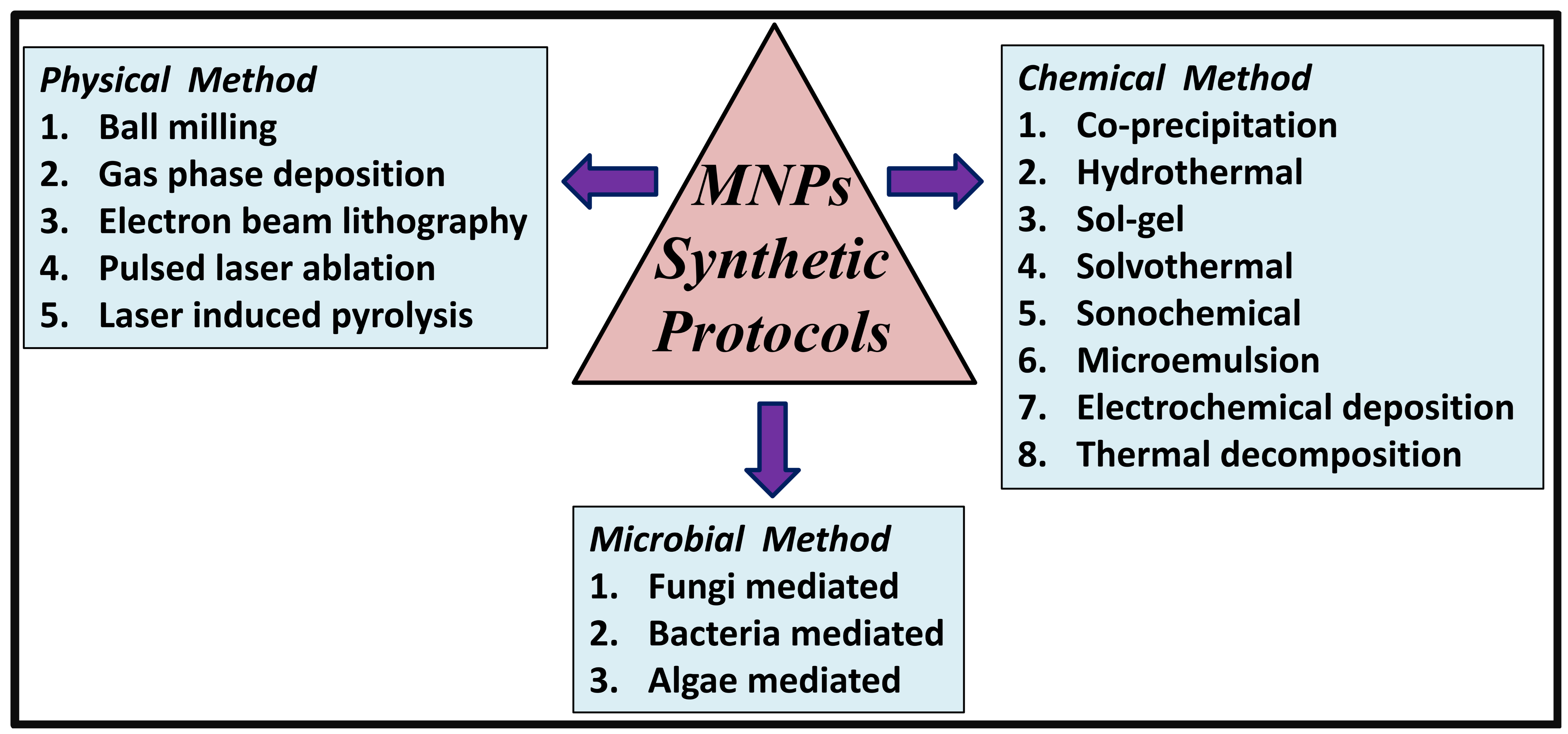
Figure 2. Synthetic Methodologies to Produce MNPs.
Physical methods typically refer to the “top-down” synthetic approaches of producing MNPs, viz. gas phase deposition[20], electron beam lithography[21], ball milling[22], pulsed laser ablation[23], laser induced pyrolysis[24], etc. Physical methods usually involve either size reduction of bulk materials to nano-sized particles or precursor condensation from gaseous/liquid phase. These techniques provide good scalability and are economically friendly. However, the obtained nano-sized particles lack uniformity of desired size and shapes[19].
Chemical method of producing MNPs basically refer to the “bottom-up” synthetic approaches viz. co-precipitation[25], hydrothermal[26], sol-gel[27], solvothermal[28], sonochemical[29], microemulsion[30], electrochemical deposition[31], thermal decomposition[32], etc. Chemical methods are the most popular protocols of preparing MNPs, as the prepared nanoparticles exhibits homogeneous chemical composition, less defects, superior short and long range ordering[33]. These protocols usually favour aqueous paths for synthesis and the obtained nanoparticles exhibits good uniformity of size and shape. The desired morphology, size, composition, and physicochemical characteristics of the MNPs could be easily achieved by the selection of appropriate synthetic approach, precursors, reaction time, pH, reaction conditions, ionic strength of media, pressure and temperature. These factors have been reported to exhibit significant effects on the shape and size of the final product. For instance, MNPs prepared through co-precipitation method using FeCl3 and FeSO4 in presence of polyvinylpyrrolidone and NaOH at 55 °C results in spherical-shaped particles of size ~9nm[34]. On the contrary, hydrothermally synthesized MNPs using FeCl3·6H2O in presence of polyethylene glycol and hydrazine at 160°C for 4h results in cubic-shaped particles of size 25 to 300nm[35]. The formation of MNPs through co-precipitation technique that comprises of mixing ferric (Fe2+) and ferrous (Fe3+) ions in a molar ratio of 1:2 in basic solution under inert atmosphere at room temperature or at elevated temperature, involves the following reactions:
Fe2++2OH-→Fe(OH)2
Fe3++3OH-→Fe(OH)3
Fe(OH)2+2Fe(OH)3→Fe3O4+4H2O
The overall reaction is:
Fe2++2Fe3++8OH-→Fe3O4+4H2O
Microbial methods or biological methods produce MNPs through fungi-mediated[36], bacteria-mediated[37], algae-mediated[38], etc. routes. These micro-organisms produce MNPs through either biologically induced or controlled mineralization modes[39]. Biologically induced mineralization mode involves different environmental factors such as temperature, pH, redox potential, pO2, pCO2, etc. to govern the production of MNPs. On the contrary, biologically controlled mineralization mode activates the microbe itself to govern MNPs synthesis. Microbial synthetic techniques are eco-friendly as they occur at ambient pressure, temperature, pH, etc. and produces stable MNPs in good yield. These techniques are highly scalable and reproducible. However, the synthesis rate is quite slow and the nanoparticles lack monodispersity[40]. To overcome these short-comings, various factors such as microbial cultivation techniques, extraction method, etc. are required to be optimized[40]. Additionally, combinatorial approach such as photo-biological techniques could be adopted. Apart from microbes, plant species and/or plants’ parts have also been used to produce MNPs[41], as they are rich source of amino acids, terpenoids, alkaloids, polysaccharides, sugars, polyphenols, proteins, carboxyl, carbonyl, etc. that acts as reducing and capping agents during MNPs synthesis.
The aforementioned synthetic protocols produce MNPs in various morphologies viz. nanorods, spherical, cube, core-shell, tube, granular, octahedron, etc. (Table 1). Prior to the environmental or biomedical applications, the synthesized MNPs are usually surface coated to bestow chemical functionalities for the attachment of bioactive molecules and to improve their aqueous dispersibility and colloidal stability. Appropriate surface coating also reduces precipitation and agglomerate formation. These surface modifications are commonly carried out through in situ coating, post-synthesis grafting or post-synthesis adsorption[19].
Table 1. Morphology and Size of MNPs Produced through Different Synthetic Protocols
Synthetic approach |
Morphology |
Size (nm)* |
Characteristics of synthetic method |
Ref. |
Spherical |
~9 |
Pros: Simple, cost-effective, particle morphology tuneable Cons: agglomerate formation Adaptable for large scale production Impurity may cause toxicity |
[34] |
|
Hydrothermal |
Cubic |
25-300 |
Pros: Homogenous particle size Cons: Sophisticated equipment required Large scale production challengeable Products are eco-friendly |
[35] |
Hydrothermal |
Nanorod |
20-30 (diameter); ~350 (length) |
|
[42] |
Co-precipitation |
Core-shell |
~ 20 |
|
[43] |
Gas phase deposition |
Granular |
~ 3 |
Pros: Eco-friendly, durable products, good purity Cons: Sophisticated equipment required Large scale production challengeable Products are eco-friendly |
[44] |
Thermal decomposition |
Octahedron |
8-~ 430 |
Pros: Cost-effective, waste management Cons: produce hazardous byproducts Large scale production little challengeable Byproducts may cause toxicity |
[45] |
Hydrothermal |
Tube |
50-100 (diameter); 200-300 (length) |
|
[46] |
Notes:*Size determined by TEM studies.
3 ENVIRONMENTAL APPLICATIONS OF MNPS
In the last few decades, the exploding industrialization and modernization have put tremendous burden on the environment. These industries primarily rely on the non-renewable energy source viz. fossil fuel, whose burning has resulted in several environmental consequences, including greenhouse effect. Extensive usage of fossil fuels has also raised concern for the search of alternative replenishable energy source. Among the plethora of work carried out by researchers to resolve these environmental issues, magnetic MNPs have been reported to be useful tool for the capture, sorption, separation and removal of toxic pollutants from the environment, owing to their eminent magnetic, chemical and optical properties (Figure 3).
|
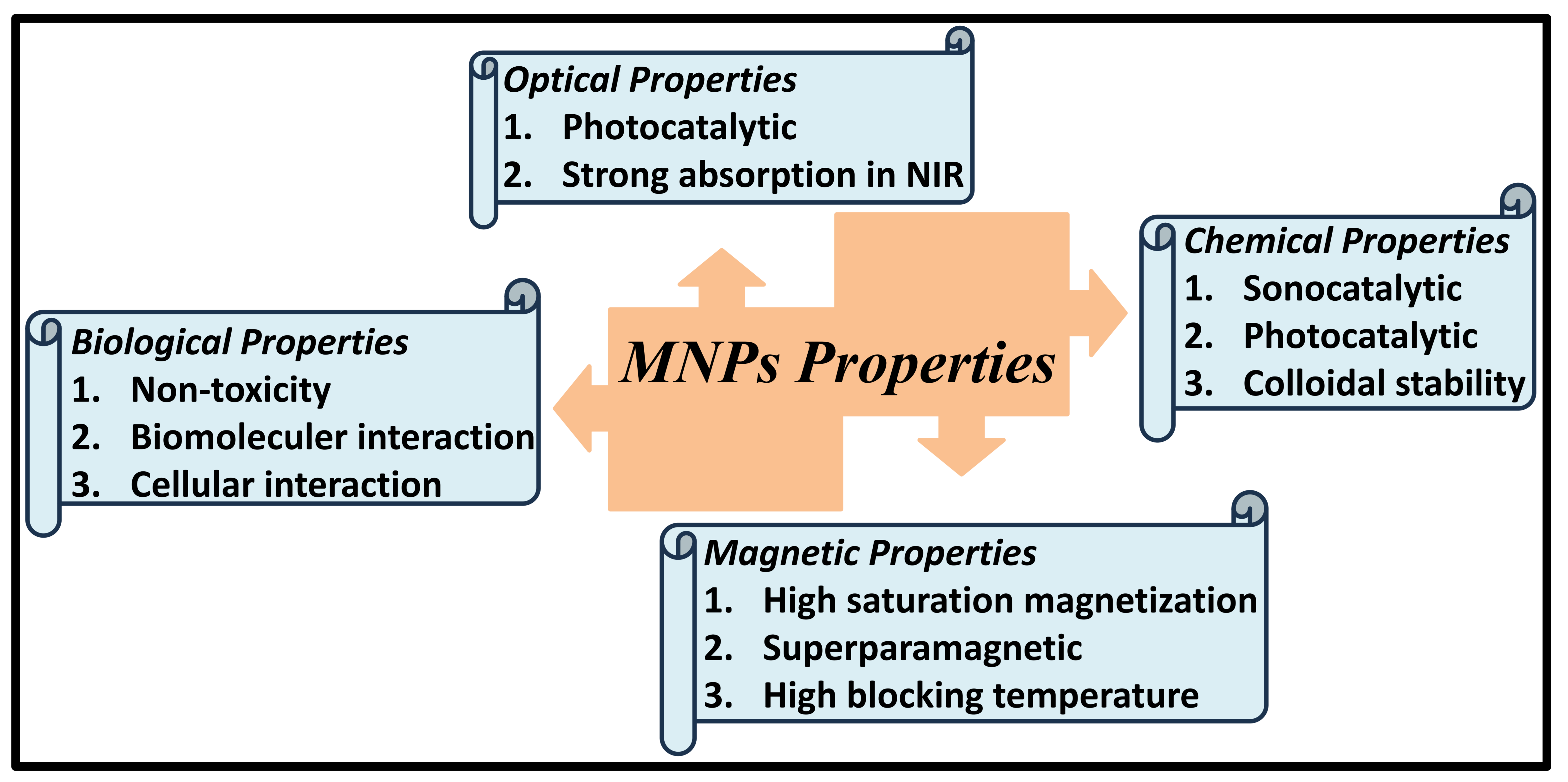
Figure 3. Few Eminent Properties of MNPs.
The extensive global production and consumption of various types of fossil fuels has led to elevated levels of greenhouse gases in the atmosphere that have resulted in numerous negative impacts on the environment such as global warming, drought, acid rains, elevation in sea levels, melting of polar ice, etc. Other activities like industrial waste, deforestation, landfills, etc., have also added enormous amount of hazardous greenhouse gases into the environment. One of the major contributors to greenhouse effect is CO2 that attributes to global warming via trapping of heat from solar radiation. Thus, seeking efficient techniques to capture and store CO2 is vital to combat the drawbacks of global warming. In this context, few innovative techniques include adsorption, absorption, membrane, and cryogenic methods[47]. The magnetic separation characteristics of MNPs offers the advantage of easy recovery and reusability (as they can be collected using external magnets) and thus are potential candidates to be utilized for the capture and storage of CO2. For instance, modified MNPs (including Fe3O4-Proline, Fe3O4-Lysine, and Fe3O4@SiO2-NH2 nanoparticles) were studied for CO2 absorption in sulfinol-M as a base solvent, where the experiments were carried out in high-pressure batch apparatus and CO2 is absorbed in sulfinol/nano-sulfinol[48]. The nanosolvents showed improved CO2 capture capacity of 6.3% and 13.36% with Fe3O4-Lysine and Fe3O4@SiO2-NH2 nanoparticles respectively at 40 bar, as compared to the base solvent. The efficiencies were observed to get further enhanced at higher pressures. In another work, adsorption of CO2 were studied using porous magnetite nanospheres (size=25.84nm; BET surface area=143.65 m2g-1), as potential CO2 sequestration reservoirs with an adsorption capacity of 0.96 mmol/g at 273 K and 1 bar of pressure[49]. On the contrary, functionalized MNPs (viz. Fe3O4-proline, Fe3O4-lysine, Fe3O4@SiO2-proline and Fe3O4@SiO2-lysine) in polypropylene hollow fiber membrane contactor have been studied for selective physical/chemical CO2 separation[50]. Results showed an eventual CO2 separation of 27.5, 57.14, 64.28, 72.8, and 96.42% for bare Fe3O4, Fe3O4-proline, Fe3O4-lysine, Fe3O4@SiO2-proline, and Fe3O4@SiO2-lysine, respectively at Ql=10mLmin−1 and Qg=100mLmin−1.
3.2 Removal of Dyes
In recent times, one of the major global challenges is inadequacy of safe and clean drinking water owing to numerous factors such as population explosion and different industrial activities. Industrialization has resulted in both huge water consumption and release of excessive waste water into different water bodies such as water drains, rivers, canals, seas and oceans. The massive quantities of hazardous industrial effluents, particularly dyes and pigments, that have been added by various industries such as paper, textile, plastics, food, paints, leather, culinary, coatings, etc. have posed a serious threat to the environment causing carcinogenic effect to aquatic and marine life. These non-biodegradable dyes change the odour and colour of water bodies, along with adversely affecting the aquatic ecosystem by consuming oxygen and escalating biochemical oxygen demand[51]. Most dyes are resistant to heat, light, other chemicals, etc. and are persistent in the environment that makes their removal or treatment from aqueous media challenging. Till date, methodologies for removal of dyes from water includes electrochemical, filtration, adsorption, coagulation, catalytic oxidation, etc. MNPs holds great potential in the removal of dyes from aqueous media due to their magnetic nature and high catalytic efficiency[52]. MNPs includes both forms of iron (Fe2+ and Fe3+) and are effective Fenton catalysts. Additionally, they have wider working pH range. Thabet and coworkers applied Box-Behnken design based on response surface methodology to study adsorptive/oxidative removal of reactive Levafix Blue dye using MNPs, where the model was used to optimize the operational parameters effect, viz. H2O2 and Fe2+/3+ doses and pH for efficient dye removal[53]. For complete dye removal of 100% in 30min or irradiance time, the optimised model (correlation coefficient, R2=94%) showed the system’s optimal pH at 2.5 with 48 and 828mg/L of MNPs and H2O2, respectively. Although, the reported optimal pH value of 2.5 is quite unrealistic, but the authors suggested that their laboratory-based experimental results could be investigated in future for application in practical and real scales. In another work, humic acid modified MNPs were prepared by in situ co-precipitation technique and studied as magnetic adsorbent for the removal of malachite green cationic dye from aqueous medium[54]. The adsorption rates follow pseudo 1st order kinetics and Freundlich isotherm model with a maximum adsorption monolayer capacity of 79.3mg/g of the sorbent. Additionally, the process was observed to be spontaneous, feasible and endothermic in nature. For reusability, the optimum desorption was attained at 0.1M of HCl.
3.3 Pesticides & Organic Pollutants
The rapid explosion in global population has raised the demand for agricultural goods leading to excessive use of pesticides and insecticides to increase crop yields. However, overuse of these persistent chemicals has led to their accumulation in water, soil, food and the environment, with a subsequent bioaccumulation in living things through the food web. Additionally, these chemicals pose considerable risk to birds, honey bees and aquatic beings. Thus, their elimination from natural sources like water and soil is urgently seeked. MNPs have been widely studied for the removal of these chemicals, due to their unique properties of superparamagnetism, strong solar light absorption, non-toxicity and ease of magnetic separation for reusability. Recently, polyacrylamide-coated MNPs were studied for the removal of imidacloprid insecticide from aqueous medium through batch adsorption experiments with spontaneous exothermic sorption process[55]. The nanoadsorbent showed adsorption capacity of 16.744mg/g and 96.71% removal of imidacloprid within 100min with pseudo-second-order kinetics, following Temkin adsorption isotherm. Another research group synthesized magnetic nanomodified activated carbon by impregnating MNPs in the activated carbon for the removal of thiamethoxam pesticide from water and sugarcane juice[56]. Results depicted highly selective chemical adsorption with pseudo-second-order kinetics (r2=0.9999), following Hill model, and sorption capacity of 42.44mg/g.
Organic pollutants such as aliphatic and polyaromatic hydrocarbons that are released into water bodies during oil spill incidents also pose a hazardous threat to the aquatic and marine ecosystems. Oil spill occurs during industrial accidents or leakage from oil tankers used in marine transport. Polyaromatic hydrocarbons, which comprises of two or more fused benzene rings, are the most toxic constituent of petroleum products with teratogenic, carcinogenic, and mutagenic effects. Although hydrophobic, they are partially water-soluble and thus are present in ground water and surface water sources. Peralta and coworkers developed magnetic silica-based nanoadsorbents for the removal of anthracene, as a polyaromatic hydrocarbon model from aqueous medium with the nanoadsorbent being easily separated from the supernatant using an external magnet[57]. The nanoadsorbent showed rapid sorption process with equilibrium attainment within 30min, could be regenerated using ethanol and reused in eight cycles with insignificant drop in anthracene removal. In another work, Patowary et al.[58] developed magnetic titania nanotubes (MNPs embedded in the nanotubes) with superhydrophobic-oleophilic nature (static water contact angle=166±1°) for the selective magnetically driven removal of spilled oil from water surfaces with maximum oil sorption capacity >1.5g/g for various oil samples. The magnetic nanotubes showed good buoyancy, high selectivity and could be reused upto five sorption cycles.
Inorganic pollutants such as heavy metals are released into the environment from different anthropogenic activities (such as mining, industrial processes, fossil fuel combination, agricultural activities, etc.) and various natural processes (such as erosion, spring waters, volcanic eruptions, bacterial activities, etc.). Long term or continuous exposure to even lower concentration of these heavy metals to living things may cause severe damage to nucleic acids and may critically disrupt various biological functions. Common remediation techniques employed for the removal of heavy metals from water and soil include ion-exchange, adsorption, reverse osmosis, chemical coagulation, precipitation, solvent extraction, filtration, etc. MNPs have been widely studied as potent candidate for the removal of heavy metals from the environment owing to their magnetic, sonocatalytic, and photocatalytic nature[59], along with other properties such as good stability, reusability and recyclability. For instance, MNPs were synthesized by co-precipitation method and studied for the removal of heavy metals viz. Cu, Cr, Cd, Pb, Ni, Fe, and Zn from soil and water samples in a remediation time <20min[60], where maximum adsorption was observed for lead with a removal efficiency of 98% and 99.6% in water and soil samples, respectively. In water samples, removal efficiency for different metals was observed to be highest at pH 4.5 with values between 63.5 and 98.3%, whereas in soil samples maximum efficiency was observed at pH 0.7 with values between 69.6 and 99.6%. As the optimum pH values of 4.5 and 0.7 are quite unrealistic, the authors suggested that the stability and removal efficiency of adsorbed products could be enhanced by modifying the MNPs with functional groups and adjusting the pH values towards 7. In another work, coal fly ash was used to synthesize MNPs of purity ~90-93% and size 30-70 nm, and subsequently used to remove toxic metals present in coal fly ash (viz. As2+, Pb2+, Cd2+, Cr6+, Cr4+, Ni2+, Co2+, Cu2+, Cu3+, etc.) from 20% w/v coal fly ash wastewater with a removal efficiency of 40-70%[61]. The MNPs could remove about nine heavy metals (i.e., Al, Cu, Cd, Cr, Co, Mn, Ni, Pb and Zn) efficiently from aqueous medium with maximum adsorption efficiency at 60min.
3.5 Pharmaceutical and Personal Care Products
The escalation in the usage of pharmaceutical and personal care products in recent years has received broad wide attention due to their contribution in the contamination of water bodies, including drinking water, sewage effluents, groundwater and surface water. Commonly detected pharmaceutical and personal care products in water sources include analgesics, antibiotics, antipyretics, immunosuppressive drugs, hormone cytostatic agents, disinfectants, etc. Most of these products are non-biodegradable and addition of these pharmaceuticals in drinking water sources cause disastrous effects to health, along with increasing drug resistance in human being. Numerous research work are, therefore, being carried out for their efficient removal or degradation from the environment. In this context, MNPs are potent candidates for the removal of these pollutants from the environment particularly as magnetic adsorbents, owing to their large surface area, high adsorption ability, stability, photocatalytic nature, and ease of recyclability. For instance, magnetite-hematite combined nanoparticles were used for the photocatalytic degradation of acetaminophen (analgesic and antipyretic drug) and caffeine (central nervous system stimulant) from wastewater[62]. The magnetic photocatalyst could remove total acetaminophen and caffeine (20ppm/150mL) using a 0.13g catalyst/L solution in 45min and 60min, respectively under UV-vis irradiation, and could be recycled for at least five experiments with no performance loss. In another work, magnetite red mud nanoparticles were studied for the removal of psychiatric drugs viz. carbamazepine, diazepam, fluoxetine, lorazepam and paroxetine from waste water[63]. The adsorption of carbamazepine (model compound) showed pseudo-second-order kinetics and Freundlich isotherm model with an adsorption capacity of 90.5mg/g in 30min at pH 6.5-7.0 and 25°C. The nanoparticles were used to treat carbamazepine from real wastewater treatment plants effluents and were observed to be reusable for five cycles. In another instance, green synthesized MNPs were studied for the removal of mixture of seven antibiotics, viz. tazobactam, piperacillin, tetracycline, sulfamethoxazole, ampicillin, trimethoprim, and erythromycin from aqueous media using Box-Behnken design approach with a removal efficiency of >90%, following pseudo-second order kinetics and Langmuir isotherm [64]. In similar work, MNPs have been studied for the removal of ciprofloxacin[65], diclofenac and venlafaxine[66].
4 BIOMEDICAL APPLICATIONS OF MNPS
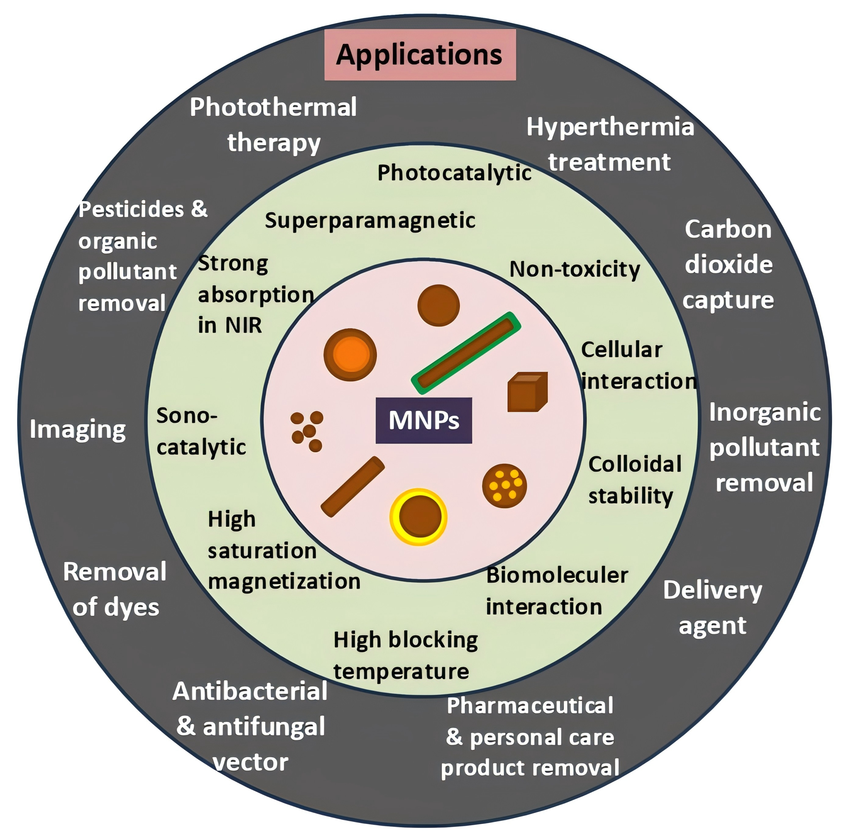
In recent decades, the advancing modernization and industrialization have posed a great threat to human health. Numerous factors such as processed food, unbalance lifestyle, pollution, and weather change have threatened human health severely[67]. These factors may cause several serious diseases, with cancer being one of the most popular diseases. Owing to the ability of MNPs to function at both molecular and cellular levels, they are considered as potent candidate for application in various biomedical fields. Other eminent properties such as non-toxicity and magnetic nature have led to the in vitro and in vivo application of MNPs in diverse areas, including delivery agents (drugs and genes), hyperthermia treatment, imaging, photothermal therapy, antibacterial/antifungal vectors, etc. (Figure 4).
|
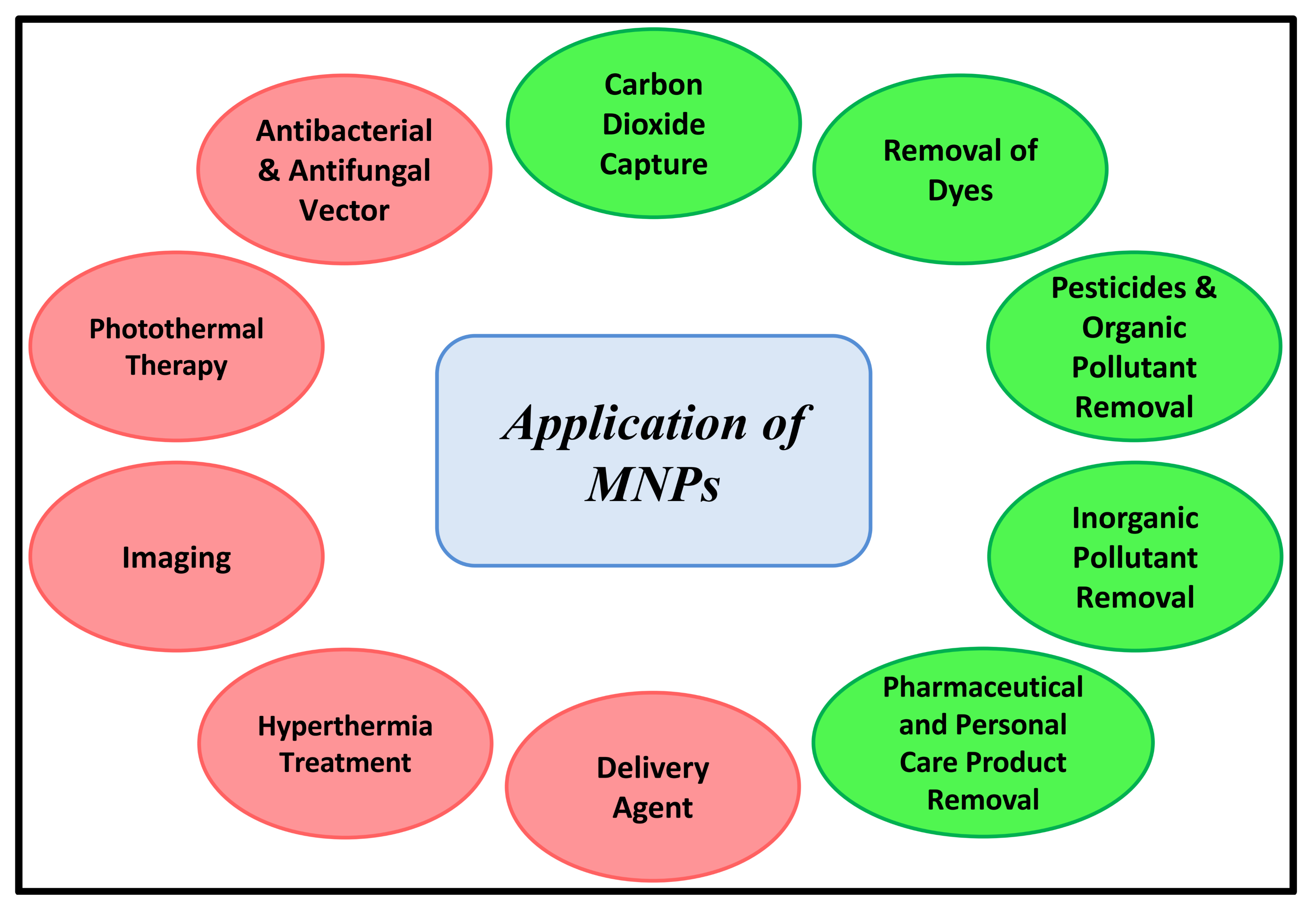
Figure 4. Common Application of MNPs in Environmental and Biomedical Fields.
4.1 Delivery Agent
Over the last few decades, tremendous advancements have been made in the development of novel therapeutics for the treatment of numerous diseases, particularly those that lacks effective treatment options. Although these therapeutics have been proved to be safe in clinical trials, they often produce numerous side effects in patients such as gastrointestinal disturbances, nausea, skin irritation, and other long-term complications. Additionally, some therapeutics such as genetic materials, large proteins, poorly soluble molecules have poor therapeutic efficacy upon administration via conventional methods such as oral administration, systemic administration or direct injections. Delivery techniques, therefore, have been observed to be a boon in the mitigation of few side effects related to unspecific tissue delivery, immune response, and rapid molecule degradation[68]. In addition, delivery methods improve the therapeutic efficacy of these therapeutic agents. MNPs have been found to be excellent magnetically-guided delivery vehicles for the targeted delivery of drugs, genes, etc. (Figure 5). For instance, luteinizing hormone releasing hormone conjugated anticancer drugs viz. prodigiosin and paclitaxel were delivered to target sites using MNPs modified microporous poly-dimethyl siloxane system for the targeted suppression of triple-negative breast cancer cells with considerably higher (p<0.05) percentage of cell growth inhibition[69]. Recently, Pd(DMEN)Cl2 complex with antitumor activity was loaded onto MNPs as targeted drug delivery system for liver cancer treatment with drug release characteristics in acidic environment of pH 4.5[70]. In vivo examinations in rat model depicted that the combination of Pd complex with MNPs showed a potential antitumor activity on liver cancer cells, with improvement in liver functions, structure and mitigation of inflammation. In similar work, MNPs have been studied as magnetic drug delivery carrier for the delivery of model anti-tumor drug curcumin[71], antibiotic drug ciprofloxacin[72], analgesic drug aceclofenac[73], etc.
|
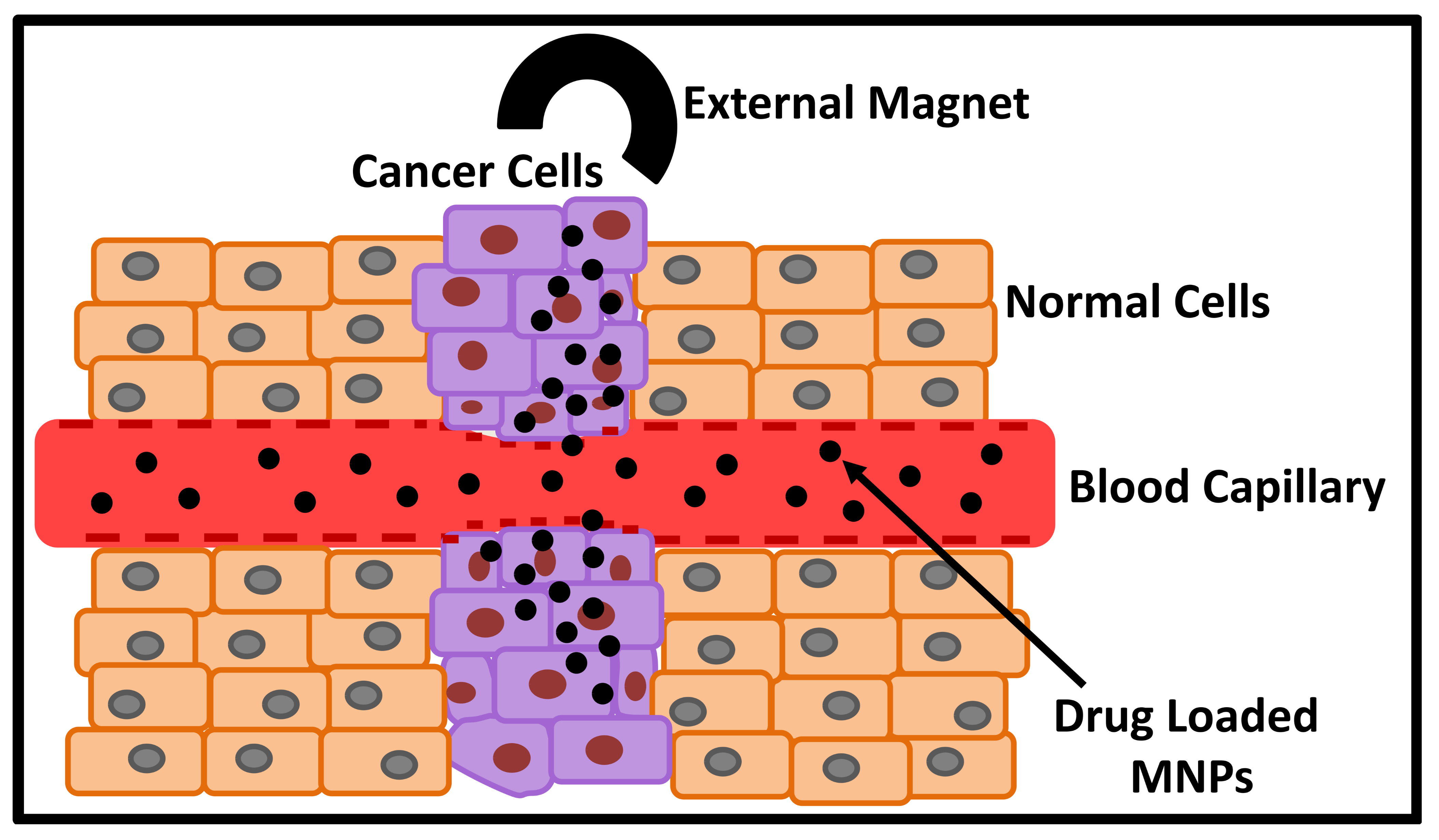
Figure 5. Magnetically Guided Drug Delivery by MNPs.
Another contemporary medical therapy for the prevention and treatment of diseases is gene therapy that encompasses the usage of genetic materials to modify the genetics underlying genome-related disorders[74]. However, the success of gene therapy is completely dependent on the selection of suitable vector for the delivery of these genetic materials as most of the therapeutic genes are unstable macromolecules[75]. Among non-viral vectors, MNPs are potent candidates for gene delivery to the target sites. For instance, transgene carrier comprising of MNPs surface-functionalized with polyethyleneimine were used for miRNA34a delivery through magnetofection as gene delivery system to downregulate programmed death-ligand 1 expression in cancer cells[76]. The magnetic carrier loaded with plasmid DNA encoding GFP depicted excellent transfection efficacy at various time points and weight ratios in MDA-MB-231 and A549 cells. Recently, green-synthesized superparamagnetic MNPs were studied for simplified nanocarrier-based genetic transformation in plant genetic engineering involving effectual DNA transfer into Catharanthus roseus leaf cells, where the delivery vehicle was loaded with the mGFP5-ER-encoding plasmid for transient gene expression in plant tissue[77].
4.2 Hyperthermia Treatment
In recent decades, cancer has emerged as one of the deadliest diseases in the world. Although, conventional monotherapies such as surgery, radiotherapy and chemotherapy show certain inhibitory effects on malignant tumors, these therapies could be harmful to normal cells around the lesion and some could lead to spread of cancer cells. Therefore, hyperthermia treatment has emerged as a novel approach for cancer treatment with minimum side effects. Hyperthermia involves the inhibition and killing of cancer cells via local hyperthermia (42-45°C), but local hyperthermia lacks targeted therapy and may cause side effects such as burns to normal tissues[78]. Thus, magnetic hyperthermia with non-invasive and targeting characteristics is an excellent hyperthermia technique that could be used as an adjuvant for radiotherapy or chemotherapy. This method uses magnetic nanoparticles such as MNPs that are conveyed or injected via the blood stream to the tumor sites, activated by an alternating current magnetic field, with subsequent thermal energy release by hysteresis losses that increases the temperature of tumor region from 37°C to 45°C[79,80]. MNPs exhibits some excellent properties such as low coercivity, high magnetic saturation, magnetic anisotropy, loop shifting, high blocking temperature, colloidal stability and biocompatibility that makes them ideal candidates for magnetic hyperthermia[80]. Darwish and coworkers prepared polyvinyl alcohol coated MNPs through ultrasonic-assisted coprecipitation method with good aqueous stability, magnetization saturation of 45.08emu/g, specific loss power of 163.81W/g (at frequency=614.4kHz; magnetic field=9.5kA/m) and excellent heating performance for potential use as magnetic hyperthermia agent for cancer therapy[81]. In another work, tumor-colonizing bacteria Escherichia coli were used as carrier for the delivery of MNPs to hypoxic tumor cores for magnetic hyperthermia-based cancer therapy that involves the incorporation of anisotropic magnetic nanocubes into Escherichia coli resulting in self-propelling nano-bacteriomagnets[82]. The nano-bacteriomagnets with specific absorption rate of 107W/g (at frequency=165kHz; amplitude=375Oe), showed 85% cell death in pancreatic cancer in in-vitro studies, while in vivo studies showed 50% tumor inhibition rate in 20 days and 100% tumor regression in 30 days. Recently, two unique structured MNPs viz. hollow spherical and nanoflower were studied for magnetic hyperthermia therapy with improved magnetothermal conversion efficiency, where the nanoflowers showed an intrinsic loss power of 6.52, while hollow spherical showed a value of 3.55[83].
4.3 Imaging
One of the challenging task in biomedical applications is the monitoring of delivery agents to the target sites/specific organs and visualizing physiological processes in the body during normal and diseased conditions. In this contrast, magnetic MNPs are ideal candidates for application as tracer agent in magnetic particle imaging (MPI) and contrasting agent in magnetic resonance imaging (MRI). MPI is an emerging, preclinical, tomographic, tracer-based, three-dimensional imaging technique which is competent in the visualization of concentration distribution of MNPs. MPI is free of ionizing radiation and gives real-time visualization, and thus could be used in vascular imaging. On the other hand, MRI is a non-invasive method that has been widely utilized in the visualization of body parts and various physiological processes of the body during diseased and normal conditions, along with the detection of degradation of delivery carriers and release of drugs. MRI agents usually comprise of T2 contrast agents which are superparamagnetic nanoparticles that shortens the transverse relaxation time, and T1 contrast agents which are paramagnetic manganese or gadolinium complexes that shortens the longitudinal relaxation time of protons[84]. T2 contrast agents results in darker images, while T1 contrast agents results in brighter images. Bente and co-workers prepared ~200nm MNPs as fast swarming microscopic units that develops into a millimeter-sized cloud with speeds of 8 mm/s, which could eventually be imaged and selectively steered using a MPI scanner[85]. In another work, (MNPs/poly(ε-caprolactone))/chitosan nanostructure was prepared via interfacial polymer disposition and coacervation and subsequently loaded with gemcitabine for potential use as tumor-targeted theranostic platform for tumor MRI and chemotherapy[86]. In vivo MRI studies on SCID mice showed good biodistribution of the nanostructures in tumor tissue samples upon using magnetic fields. The relaxivity values of the hybrid MNPs (with r1=0.3, r2=70.6mM-1s-1; r2/r1=~235) were found to be similar to that for Ferumoxytol contrast agent. Table 2 summarizes the magnetic characteristics of MNPs that have been studied for hyperthermia, MPI and MRI applications.
Table 2. Magnetic Characteristics of MNPs Studied for Hyperthermia, MPI and MRI Applications
Synthesis method |
Saturation magnetization |
Specific absorption rate (SAR); frequency; amplitude |
Hysteresis |
Size (nm) |
Zeta potential (mV) |
Relaxivity; (r₁/r₂) |
Application |
Ref. |
Co-precipitation |
- |
- |
67.9±4.8 (TEM) 193.4±7.1 (hydrodynamic) |
-37.0±2.1 |
- |
Hyperthermia therapy |
[82] |
|
Solvothermal |
at 150mT |
- |
Dynamic 25kHz, 12mT |
200 (SEM) |
- |
- |
MPI |
[85] |
Thermal decomposition |
~30emu/g |
- |
- |
2.3 (TEM) |
- |
r1=6 r2= 24.2mM-1s-1; r2/r1=4 |
MRI and angiography |
[84] |
Co-precipitation |
11.34±0.81Am2/kg |
- |
Susceptibility 0.156±0.026)×10-3m3/kg |
~308 (hydrodynamic) |
28 |
MRI and chemotherapy |
[86] |
|
Chemical precipitation |
47.56emu/g |
- |
Hc=33Oe |
15-30 (TEM) |
- |
- |
Hyperthermia therapy |
[78] |
Co-precipitation |
52.04emu/g |
33 W/g at 9mg/mL; Frequency=92kHz; Amplitude=10kA/m |
Hc=7.35Oe |
3-40 (TEM) 16.6 (hydrodynamic) |
- |
- |
Hyperthermia therapy |
[80] |
Co-precipitation |
76Am2/kg |
170W/g; Frequency=100kHz; Amplitude=48kA/m |
Hc=1kA/m |
95±18 (hydrodynamic) |
-26.1±1.4 |
- |
Hyperthermia therapy |
[79] |
4.4 Photothermal Therapy
In recent years, photothermal therapy has emerged as a promising therapeutic technique for the destruction of malignant tissues. Upon using photothermal converters, light energy is effectively converted into heat energy. Generally, nanoparticles are loaded into malignant tissues and subsequently irradiated with near-infrared (NIR) laser to produce heat that ultimately destroys the malignant tissues. This technique exhibits numerous advantages including precise spatial temporal selectivity, high specificity, and minimal invasiveness[87]. Additionally, photothermal therapy modality doesn’t need oxygen and could be carried out using longer wavelength radiation (700-1000nm) and thus less harmful to normal cells[87]. MNPs have been widely studied as efficient photothermal converters in photothermal therapy due to their strong absorption in the NIR window[88]. Sun and co-workers synthesized MNPs via chemical method and studied them for photothermal therapy and immunotherapy in the treatment of head and neck squamous cell carcinoma[89]. In vitro cellular and in vivo animal experiments showed that photothermal properties of MNPs directly triggers immunogenic cell death and improves immunogenicity of the tumor micromilieu, along with regulating the expression of tumor-associated macrophages, up-regulation of CD86 and downregulation of CD206 for the inhibition of tumor growth. Recently, MNPs were co-doped with 2% Co and 40% Zn divalent ions and studied for hyperthermia and photothermal therapy using 3D alveolar reconstructed A549 lung cancer tissue model[90]. It was observed that photothermal therapy exhibits improved heat generation response as compared to hyperthermia with an external photothermal conversion efficiency of ∼1.3gL-1cm-1 at 808nm.
4.5 Antibacterial/Antifungal Vectors
Microbial infections viz. bacterial and fungal is one of the frequent reasons for hospitalization worldwide, whereas nosocomial infections commonly trigger the rise in morbidity and mortality, particularly within healthcare facilities[91]. Additionally, patients who receives antibiotic or antifungal therapies harbour a huge number of antibiotic or antifungal resistant microbes in their intestinal tracts which are eventually excreted in faeces and contaminates the environment through feacal materials[92]. As a result, antibiotic or antifungal resistant microbes have been detected in sewage, rivers and other polluted water bodies. Recently, MNPs have been investigated as potent antimicrobial agent to cope with the threat of antimicrobial resistance. MNPs could be managed with external magnetic field, possess low internal diffusion resistance and could destroy microbes via interference with thiol groups of these microorganisms present at their respiratory base[91]. Consequently, MNPs have been studied as antimicrobial vector against Escherichia coli[93-95], Aspergillus niger[93-95], Staphylococcus aureus[93-95], Baccilis subtilis[95], Saccharomyces cerevisiae[95], Candida albicans[95], Trichoderma spp.[95], Aspergillus fumigatus[96], Fusarium verticilliodes[97], Aspergillus flavus[97], Alternaria alternata[97], Aeromonas sobria[98], etc. Dabagh and co-workers green synthesized MNPs using lemon fruit extract, which were then capped with silica layer with subsequent decoration with silver nanoparticles[99]. The synthesized nanoparticles were then studied as antibacterial agent against Escherichia coli and Staphylococcus aureus, where excellent results were observed even at low concentrations (≤200μg/mL). In another work, MNPs were synthesized through a green method using spinach as precursor and extracts of pomegranate juice, pomegranate peel, white vinegar, aloe vera peel, black coffee, and aspirin as reducing/stabilizing agents for tuning the properties of MNPs[100]. The green-synthesized MNPs were subsequently studied as antifungal vector for the inhibition of mycelial growth of Fusarium oxysporum f. sp. lycopersici via both in vitro and in vivo assays with considerable reduction in disease severity and incidence. Table 3 summarizes various reported hybrid MNPs that have been studied for different biomedical applications, whereas Table 4 summarizes the characteristics of various MNPs that have been studied for different environmental and biomedical applications.
Table 3. Summary of Various Hybrid MNPs and Their Potential Biomedical Applications
Hybrid MNPs |
Biomedical Applications |
Ref. |
Poly-dimethyl siloxane functionalized MNPs |
Prodigiosin and paclitaxel delivery in breast cancer treatment |
[69] |
MNPs and Pd(DMEN)Cl2 complex |
Liver cancer treatment |
[70] |
Polyethyleneimine functionalized MNPs |
miRNA34a delivery in cancer treatment |
[76] |
Polyvinyl alcohol functionalized MNPs |
Magnetic hyperthermia for cancer therapy |
[81] |
Co and Zn co-doped MNPs |
Hyperthermia and photothermal therapy in lung cancer treatment |
[90] |
MNPs capped with silica and silver nanoparticles |
Antibacterial agent |
[99] |
MNPs encapsulated alginate beads |
Delivery of analgesic drug aceclofenac |
[73] |
SiO2@MNPs incorporated Escherichia coli |
Nano-bacteriomagnets for magnetic hyperthermia in cancer therapy |
[82] |
Mn doped MNPs |
MPI |
[101] |
Polyelectrolyte MNPs capsules |
Magnetic resonance imaging |
[102] |
NaYF4:Yb,Er@SiO2@MNPs |
Optical imaging and photothermal therapy |
[103] |
Table 4. Characteristics of Various MNPs Studied for Environmental and Biomedical Applications
Synthesis Method |
ParticlE size (nm)* |
Saturation Magnetization (emu/g) |
Crystallinity |
Coating Materials |
Applications |
Ref. |
Co-precipitation |
29.2 |
60 |
NA |
No coating |
Methylene blue dye removal |
[52] |
Co-precipitation |
21.25 |
59.77 |
Crystalline |
Polyacrylamide |
Imidacloprid insecticide removal |
[55] |
Hydrothermal |
84-109 |
18.6 |
Crystalline |
Silica, 3-(trimethoxysilyl)propyl-octadecyldimethyl-ammonium chloride, trimethoxyphenylsilane |
Removal of ibuprofen, diclofenac, carbamazepine, polyaromatic hydrocarbons |
[57] |
Co-precipitation |
10-30 |
~0.11 |
Polycrystalline |
None |
Removal of coal fly ash |
[61] |
Green synthesis using E. globulus |
2.34±0.53 and 4.17±1.22 |
1.5 and 2.1 |
Crystalline |
None |
Removal of Cr(VI), Cd, and Pb |
[104] |
Co-precipitation |
1.3-50 (pore size) |
22.5 and 32.9 |
NA |
Activated carbons from Eucalyptus globulus |
Removal of diclofenac and venlafaxine |
[66] |
Chemical method |
9.8±1.5 |
37.6 |
Crystalline |
Activated carbon from Lemna minor |
Removal of ciprofloxacin |
[105] |
Co-precipitation |
1.1×1.4 mm (beads size) |
0.49-0.64 |
NA |
Sodium alginate beads |
Delivery of aceclofenac |
[73] |
Chemical |
70 length (nanorods) |
44.79 |
Crystalline |
Polyethyleneimine |
Delivery of miRNA34a |
[76] |
Bacteria-mediated |
20-30 |
0.34 |
Crystalline |
None |
Hyperthermia therapy |
[106] |
Hydrothermal |
10±3 |
68.1 |
Crystalline |
None (Zn & Co doped) |
Lung cancer photothermal therapy |
[90] |
Co-precipitation |
NA |
11.3±0.8 |
NA |
Poly(ε-caprolactone) and chitosan |
Magnetic resonance imaging and gemcitabine delivery |
[86] |
Green synthesis using spinach |
20 |
19.3 |
Crystalline |
None |
Anti-fungal against Fusarium oxysporum |
[100] |
Co-precipitation |
10 |
45 |
Crystalline |
Multipotent antioxidant |
Anti-bacterial and anti-fungal |
[95] |
Notes: *Particle size determined by TEM/SEM studies.
5 ENVIRONMENTAL FATE AND TOXICOLOGY OF MNPS
The outstanding magnetic properties of MNPs has encouraged their widespread application in diverse environmental and biomedical areas owing to their ability to be guided/navigated using external magnets. Nevertheless, the potential toxicity of MNPs is still less explored. In environmental pollution remediation applications, the MNPs could be restored and collected from the affected sites using external magnets, thereby reducing their potential toxicity in the environment. However, for biomedical applications, MNPs must be non-toxic and biocompatible as they may degrade in the body and degraded products might interact within the body. MNPs of size >200nm are known to be retained by spleen through mechanical filtration, while those of size <10nm are removed via renal clearance. Thus, MNPs of size 10-100nm are considered optimum for administration in specific biomedical applications. The biodistribution, interaction, metabolism, probable toxicity, and internalization mechanism inside cells and organs of MNPs depend entirely on their surface modifications, dosage, concentration, size and shape[107]. Possible toxicity of MNPs includes ulceration, inflammation, decline in viability, decrease in growth rate, and triggering of neurobehavioral alterations. For clinical application of MNPs, the amount of dose and concentration needs to be tested in in vitro models for assessing the possible toxicity, before further testing in in vivo platforms for allowing correlations between findings. For instance, the in vitro study of spherical shaped MNPs of size 72.6±0.6nm demonstrate toxicity in THP-1 cells at a concentration of 25µg/ml, while in vivo studies in rat model at 8 weeks of age showed apoptosis of circulating erythrocytes at a concentration of 12mg/kg[108]. Thus, a lot of factors affecting toxicity such as concentration, dosage, surface chemistry, time, interaction medium, cell types, internalization modes, etc. needs to be elaborately studied to properly understand the fundamental mechanism before the usage of MNPs in healthcare.
6 CONCLUSION
MNPs are evolving materials that have drawn significant consideration from researchers and academics in diverse industries. The outstanding properties of excellent magnetic nature, large surface area to volume ratio, controllable morphology, non-toxicity, etc. have encouraged their potential application in diverse environmental pollution remediation and biomedical fields. In environmental applications, the use of MNPs as adsorbents, absorbents and membranes to capture and store CO2 provides a convenient approach to combat the drawbacks of global warming. The high catalytic efficiency, magnetic nature, stability, and reusability enable MNPs to be utilized in the removal of inorganic pollutants and dyes from aqueous media. Additionally, MNPs show good sorption characteristics that encouraged their potential use in the removal of pesticides, organic pollutants, pharmaceutical and personal care products. MNPs offers the advantage to be collected and restored from affected sites using external magnets which probably reduces their potential toxicity in the environment. However, application of these techniques is still limited in the early stage. Enormous research work is still needed in this field to address the uncertainties over environmental fate, performance in real environmental conditions, and toxicity of these MNPs and their health impacts before their widespread application. Additionally, the scalability of these MNPs needs to be assessed for their large-scale production and real-field application.
In biomedical applications, MNPs (size 10-100nm) have notably emerged in various formulations as therapeutics and imaging agent in the recent decades, owing to their non-toxicity, targeting ability, magnetic behaviour, hysteresis nature, and strong absorption in NIR window. However, further research is still required for understanding the physicochemical characteristics of MNPs and their in-vivo behaviour for higher clinical translation prospects and commercialize them as a product in medical domain. Toxicity concerns for adoption of MNPs in clinical treatments could be solved through various in vitro and in vivo studies, where toxicity could arise from toxicity of the precursors, normal functioning of the body cycle inside an organism and disturbances in the ongoing cellular mechanisms[107]. For better understanding of underlying mechanism, multi-omics studies using metabolomics, genomics and proteomics must be carried out to assess the toxicity of these MNPs at precise levels before their adoption in clinical treatments. From the perspective of commercial transformation, numerous characteristics needs to be considered while selecting MNPs for biomedical applications such as synthesis technique, purification, morphology, surface charge, saturation magnetization, colloidal stability, drug/gene loading capacity and release nature, etc. We expect that this review confers an incentive to researchers to search for novel insights into MNPs for diverse environmental and biomedical applications. Additionally, other magnetic materials of iron viz. ferrites[109,110], doped MNPs[111], nanocomposites[112-116], pyrites[117], etc. could also be explored for diverse environmental and biomedical applications, which also forms the future scope of the present review study.
Acknowledgements
Not applicable.
Conflicts of Interest
The authors declared no conflict of interest.
Data Availability
No additional data are available.
Copyright Permissions
Copyright © 2026 The Author(s). Published by Innovation Forever Publishing Group Limited. This open-access article is licensed under a Creative Commons Attribution 4.0 International License (https://creativecommons.org/licenses/by/4.0), which permits unrestricted use, sharing, adaptation, distribution, and reproduction in any medium, provided the original work is properly cited.
Author Contribution
Kalita H wrote the original draft, and Patowary M conducted the review and editing.
Abbreviation List
MNPs, Magnetite nanoparticles
MPI, Magnetic particle imaging
MRI, Magnetic resonance imaging
NIR, Near-infrared
References
[1] Lai L, Xie Q, Chi L et al. Adsorption of phosphate from water by easily separable Fe3O4@SiO2 core/shell magnetic nanoparticles functionalized with hydrous lanthanum oxide. J Colloid Interface Sci, 2016; 465: 76-82.[DOI]
[2] Zhang Y, Ni S, Wang X et al. Ultrafast adsorption of heavy metal ions onto functionalized lignin-based hybrid magnetic nanoparticles. Chem Eng J, 2019; 372: 82-91.[DOI]
[3] Ko S, Kim ES, Park S et al. Amine functionalized magnetic nanoparticles for removal of oil droplets from produced water and accelerated magnetic separation. J Nanopart Res, 2017; 19: 132.[DOI]
[4] Wang X, Shi Y, Graff RW et al. Developing recyclable pH-responsive magnetic nanoparticles for oil–water separation. Polymer, 2015; 72: 361-367.[DOI]
[5] Gong Y, Gai L, Tang J et al. Reduction of Cr(VI) in simulated groundwater by FeS-coated iron magnetic nanoparticles. Sci Total Environ, 2017; 595: 743-751.[DOI]
[6] Guibert C, Dupuis V, Peyre V et al. Hyperthermia of magnetic nanoparticles: experimental study of the role of aggregation. J Phys Chem C, 2015; 119: 28148-28154.[DOI]
[7] Ruta S, Chantrell R, Hovorka, O. Unified model of hyperthermia via hysteresis heating in systems of interacting magnetic nanoparticles. Sci Rep, 2015; 5: 9090.[DOI]
[8] Ayubi M, Karimi M, Abdpour S et al. Magnetic nanoparticles decorated with PEGylated curcumin as dual targeted drug delivery: Synthesis, toxicity and biocompatibility study. Mat Sci Eng C, 2019; 104: 109810.[DOI]
[9] Chowdhuri AR, Singh T, Ghosh SK et al. Carbon dots embedded magnetic nanoparticles@chitosan@metal organic framework as a nanoprobe for pH sensitive targeted anticancer drug delivery. ACS Appl Mater Interfaces, 2016; 8: 16573-16583.[DOI]
[10] Yang C, Lin G, Zhu C et al. Metalla-aromatic loaded magnetic nanoparticles for MRI/photoacoustic imaging-guided cancer phototherapy. J Mater Chem B, 2018; 6: 2528-2535.[DOI]
[11] Yang HM, Park CW, Park S et al. Cross-linked magnetic nanoparticles with a biocompatible amide bond for cancer-targeted dual optical/magnetic resonance imaging. Colloids Surf B, 2018; 161: 183-191.[DOI]
[12] Mirković M, Radović M, Stanković D et al. 99mTc–bisphosphonate–coated magnetic nanoparticles as potential theranostic nanoagent. Mat Sci Eng C, 2019; 102: 124-133.[DOI]
[13] Sheng Y, Li S, Duan Z et al. Fluorescent magnetic nanoparticles as minimally-invasive multi-functional theranostic platform for fluorescence imaging, MRI and magnetic hyperthermia. Mater Chem Phys, 2018; 204: 388-396.[DOI]
[14] Chen Y, Xianyu Y, Dong M et al. Cascade reaction-mediated assembly of magnetic/silver nanoparticles for amplified magnetic biosensing. Anal Chem, 2018; 90: 6906-6912.[DOI]
[15] Cao L, Zhang Q, Dai H et al. Separation/concentration‐signal‐amplification in‐one method based on electrochemical conversion of magnetic nanoparticles for electrochemical biosensing. Electroanalysis, 2018; 30: 517-524.[DOI]
[16] Hedayatnasab Z, Abnisa F, Daud WMAW. Review on magnetic nanoparticles for magnetic nanofluid hyperthermia application. Mater Des, 2017; 123: 174-196.[DOI]
[17] Ashraf M, Khan I, Usman M et al. Hematite and magnetite nanostructures for green and sustainable energy harnessing and environmental pollution control: a review. Chem Res Toxicol, 2020; 33: 1292-1311.[DOI]
[18] Yew YP, Shameli K, Miyake M et al. Green biosynthesis of superparamagnetic magnetite Fe3O4 nanoparticles and biomedical applications in targeted anticancer drug delivery system: A review. Arab J Chem, 2020; 13: 2287-2308.[DOI]
[19] Mohammed L, Gomaa HG, Ragab D et al. Magnetic nanoparticles for environmental and biomedical applications: A review. Particuol, 2017; 30: 1-14.[DOI]
[20] Reyes-Reyes M, Hernández-Arriaga D, López-Sandoval R. Carbon-coated hexagonal magnetite nanoflakes production by spray CVD of alcohols in mixture with water. Mater Res Express, 2014; 1: 045607.[DOI]
[21] Guillaume A, Scholtyssek JM, Lak A, et al. Magnetorelaxometry of few Fe3O4 nanoparticles at 77 K employing a self-compensated SQUID magnetometer. J Magn Magn Mater, 2016; 408: 46-50.[DOI]
[22] Prabhu K, Aralekallu S, Sajjan VA et al. Non-precious cobalt phthalocyanine-embedded iron ore electrocatalysts for hydrogen evolution reactions. Sustain Energ Fuels, 2021; 5: 1448-1457.[DOI]
[23] Svetlichnyi VA, Shabalina AV, Lapin IN et al. Comparative study of magnetite nanoparticles obtained by pulsed laser ablation in water and air. Appl Surf Sci, 2019; 467-468: 402-410.[DOI]
[24] Morjan I, Alexandrescu R, Dumitrache F et al. Iron oxide-based nanoparticles with different mean sizes obtained by the laser pyrolysis: structural and magnetic properties. J Nanosci Nanotechnol, 2010; 10: 1223-1234.[DOI]
[25] Radoń A, Drygała A, Hawełek L et al. Structure and optical properties of Fe3O4 nanoparticles synthesized by co-precipitation method with different organic modifiers. Mater Charact, 2017; 131: 148-156.[DOI]
[26] Long LQ, Hue TTB, Hoan NX et al. Growth mechanism and stability of magnetite nanoparticles synthesized by the hydrothermal method. J Nanosci Nanotechnol, 2016; 16: 7373-7379.[DOI]
[27] Shi S, Xu C, Wang X et al. Electrospinning fabrication of flexible Fe3O4 fibers by sol-gel method with high saturation magnetization for heavy metal adsorption. Mater Des, 2020; 186: 108298.[DOI]
[28] Fotukian SM, Barati A, Soleymani M et al. Solvothermal synthesis of CuFe2O4 and Fe3O4 nanoparticles with high heating efficiency for magnetic hyperthermia application. J Alloys Compd, 2020; 816: 152548.[DOI]
[29] Neto DMA, Freire RM, Gallo J et al. Rapid sonochemical approach produces functionalized Fe3O4 nanoparticles with excellent magnetic, colloidal, and relaxivity properties for MRI application. J Phys Chem C, 2017; 121: 24206-24222.[DOI]
[30] Asab G, Zereffa EA, Seghne TA. Synthesis of silica-coated Fe3O4 nanoparticles by microemulsion method: characterization and evaluation of antimicrobial activity. Int J Biomater, 2020; 2020: 4783612.[DOI]
[31] Karimzadeh I, Dizaji HR, Aghazadeh M. Preparation, characterization and PEGylation of superparamagnetic Fe3O4 nanoparticles from ethanol medium via cathodic electrochemical deposition (CED) method. Mater Res Express, 2016; 3: 095022.[DOI]
[32] Patsula V, Kosinová L, Lovrić M et al. Superparamagnetic Fe3O4 nanoparticles: synthesis by thermal decomposition of iron(III) glucuronate and application in magnetic resonance imaging. ACS Appl Mater Interfaces, 2016; 8: 7238-7247.[DOI]
[33] Wang Y, Xia Y. Bottom-Up and Top-Down Approaches to the Synthesis of Monodispersed Spherical Colloids of Low Melting-Point Metals. Nano Lett, 2004; 4: 2047-2050.[DOI]
[34] Radoń A, Łukowiec D, Kremzer M et al. Electrical conduction mechanism and dielectric properties of spherical shaped Fe3O4 nanoparticles synthesized by co-precipitation method. Materials, 2018; 11: 735.[DOI]
[35] Torres-Gómez N, Nava O, Argueta-Figueroa L et al. Shape tuning of magnetite nanoparticles obtained by hydrothermal synthesis: effect of temperature. J Nanomater, 2019; 2019: 7921273.[DOI]
[36] Abdeen M, Sabry S, Ghozlan H et al. Microbial-physical synthesis of Fe and Fe3O4 magnetic nanoparticles using aspergillus niger YESM1 and supercritical condition of ethanol. J Nanomater, 2016; 2016: 9174891.[DOI]
[37] Kim Y, Roh Y. Microbial synthesis and characterization of superparamagnetic Zn-substituted magnetite nanoparticles. J Nanosci Nanotechnol, 2015; 15: 6129-6132.[DOI]
[38] Salem DMSA, Ismail MM, Aly-Eldeen MA. Biogenic synthesis and antimicrobial potency of iron oxide (Fe3O4) nanoparticles using algae harvested from the Mediterranean Sea, Egypt. Egypt J Aquat Res, 2019; 45: 197-204.[DOI]
[39] Revati AK, Pandey BD. Microbial synthesis of iron-based nanomaterials-A review. Bull Mater Sci, 2011; 34: 191-198.[DOI]
[40] Narayanan KB, Sakthivel N. Biological synthesis of metal nanoparticles by microbes. Adv Colloid Interface Sci, 2010; 156: 1-13.[DOI]
[41] Ali I, Peng C, Naz I et al. Phytogenic magnetic nanoparticles for wastewater treatment: a review. RSC Adv, 2017; 7: 40158.[DOI]
[42] Attallah OA, Girgis E, Abdel-Mottaleb MMSA. Synthesis of non-aggregated nicotinic acid coated magnetite nanorods via hydrothermal technique. J Magn Magn Mater, 2016; 399: 58-63.[DOI]
[43] Sandoval SS, Santibanez D, Riveros A et al. Synthesis and characterization of magnetite/gold core shell nanoparticles stabilized with a β-cyclodextrin nanosponge to develop a magneto-plasmonic system. Magnetochemistry, 2023; 9: 202.[DOI]
[44] Jiang W, McCloy JS, Lea AS et al. Magnetization and susceptibility of ion-irradiated granular magnetite films. Phys Rev B, 2011; 83: 134435.[DOI]
[45] Li L, Yang Y, Ding J et al. Synthesis of magnetite nanooctahedra and their magnetic field-induced two-/three-dimensional superstructure. Chem Mater, 2010; 22: 3183-3191.[DOI]
[46] Jazirehpour M, Ebrahimi SAS. Synthesis of magnetite nanostructures with complex morphologies and effect of these morphologies on magnetic and electromagnetic properties. Ceram Int, 2016; 42: 16512-16520.[DOI]
[47] Zandahvifard MJ, Elhambakhsh A, Ghasemi MN et al. Effect of modified Fe3O4 magnetic NPs on the absorption capacity of CO2 in water, wettability alteration of carbonate rock surface, and water-oil interfacial tension for oilfield applications. Ind Eng Chem Res, 2021; 60: 3421-3434.[DOI]
[48] Elhambakhsh A, Keshavarz P. Enhanced CO2 capture efficiency applying amine-based nano magnetite/sulfinol-M nano solvents at high pressures. Environ Sci Pollut Res, 2020; 28: 3455-3464.[DOI]
[49] El-Desouky MG, El-Bindary AA, El-Bindary MA. Low-temperature adsorption study of carbon dioxide on porous magnetite nanospheres iron oxide. Biointerface Res Appl Chem, 2021; 12: 6252-6268.[DOI]
[50] Rasaie M, Elhambakhsh A, Eskandari M et al. Highly selective physical/chemical CO2 separation by functionalized Fe3O4 nanoparticles in hollow fiber membrane contactors: experimental and modeling approaches. Energy Fuels, 2022; 36: 4456-4469.[DOI]
[51] Abuessawy A, Fouda A, Abdel‑Rahman AAH et al. A new modified heterocyclic‑magnetite chitosan nanocomposite for efficient alizarin red dye removal: adsorption analysis and antibacterial activity. J Polym Environ, 2024; 32: 826-841.[DOI]
[52] Aly ST, Saed A, Mahmoud A et al. Preparation of magnetite nanoparticles and their application in the removal of methylene blue dye from wastewater. Sci Rep, 2024; 14: 20100.[DOI]
[53] Thabet RH, Fouad MK, Ali IA et al. Magnetite-based nanoparticles as an efficient hybrid heterogeneous adsorption/oxidation process for reactive textile dye removal from wastewater matrix. Int J Environ Anal Chem, 2023; 103: 2636-2658.[DOI]
[54] Abate GY, Alene AN, Habte AT et al. Adsorptive removal of basic green dye from aqueous solution using humic acid modified magnetite nanoparticles: kinetics, equilibrium and thermodynamic studies. J Polym Environ, 2021; 29: 967-984.[DOI]
[55] Rani K, Kumar R, Singh D et al. Adsorption studies of imidacloprid from aqueous solution using polyacrylamide coated magnetite nanoparticles as a potential nanoadsorbent. Biointerface Res Appl Chem, 2024; 14: 88.[DOI]
[56] de Freitas DA, Barbosa JA, Labuto G et al. Removal of the pesticide thiamethoxam from sugarcane juice by magnetic nanomodified activated carbon. Environ Sci Pollut Res, 2022; 29: 79855-79865.[DOI]
[57] Peralta ME, M´artire DO, Moreno MS et al. Versatile nanoadsorbents based on magnetic mesostructured silica nanoparticles with tailored surface properties for organic pollutants removal. J Environ Chem Eng, 2021; 9: 104841.[DOI]
[58] Patowary M, Ananthakrishnan R, Pathak K. Effective oil removal from water by magnetically driven superhydrophobic and oleophilic magnetic titania nanotubes. Environ Sci Pollut Res, 2017; 24: 18063-18072.[DOI]
[59] Lankathilaka KPW, de Silva RM, Mantilaka MMMGPG et al. Magnetite nanoparticles incorporated porous kaolin as a superior heavy metal sorbent for water purification. GSD, 2021; 14: 100606.[DOI]
[60] Singh A, Chaudhary S, Dehiya BS. Fast removal of heavy metals from water and soil samples using magnetic Fe3O4 nanoparticles. Environ Sci Pollut Res, 2021; 28: 3942-3952.[DOI]
[61] Yadav VK, Amari A, Gacem A et al. Treatment of fly-ash-contaminated wastewater loaded with heavy metals by using fly-ash-synthesized iron oxide nanoparticles. Water, 2023; 15: 908.[DOI]
[62] Fernandes TA, Mendo SG, Ferreira LP et al. Photocatalytic degradation of acetaminophen and caffeine using magnetite-hematite combined nanoparticles: kinetics and mechanisms. Environ Sci Pollut Res, 2021; 28: 17228-17243.[DOI]
[63] Aydın S, Bedük F, Ulvi A et al. Simple and effective removal of psychiatric pharmaceuticals from wastewater treatment plant effluents by magnetite red mud nanoparticles. Sci Total Environ, 2021; 784: 147174.[DOI]
[64] Stan M, Lung I, Soran ML et al. Removal of antibiotics from aqueous solutions by green synthesized magnetite nanoparticles with selected agro-waste extracts. Process Saf Environ Prot, 2017; 107: 357-372.[DOI]
[65] Salari M. Optimization by Box-Behnken design and synthesis of magnetite nanoparticles for removal of the antibiotic from an aqueous phase. Adsorp Sci Technol, 2022; 2022: 1267460.[DOI]
[66] Rocha LS, Sousa EML, Pereira D et al. Sustainable and recoverable waste-based magnetic nanocomposites used for the removal of pharmaceuticals from wastewater. Chem Eng J, 2021; 426: 129974.[DOI]
[67] Yew YP, Shameli K, Miyake M et al. Green biosynthesis of superparamagnetic magnetite Fe3O4 nanoparticles and biomedical applications in targeted anticancer drug delivery system: A review. Arab J Chem, 2020; 13: 2287-2308.[DOI]
[68] Arango D, Cifuentes J, Puentes PR et al. Tailoring magnetite-nanoparticle-based nanocarriers for gene delivery: exploiting CRISPRa potential in reducing conditions. Nanomaterials, 2023; 13: 1782.[DOI]
[69] Eluu SC, Obayemi JD, Yiporo D et al. Luteinizing hormone-releasing hormone (LHRH)-conjugated cancer drug delivery from magnetite nanoparticle-modified microporous poly-di-methyl-siloxane (PDMS) systems for the targeted treatment of triple negative breast cancer cells. J Funct Biomater, 2024; 15: 209.[DOI]
[70] El-Sayed SAM, Fouad GI, Beherei HH et al. Palladium complex-loaded magnetite nanoparticles as drug delivery systems for targeted liver cancer therapy. Pharmaceutics, 2025; 17: 1033.[DOI]
[71] Kalita H, Rajput S, Kumar BNP et al. Fe3O4@zirconium phosphate core-shell nanoparticles for pH-sensitive and magnetically guided drug delivery applications. RSC Adv, 2016; 6: 21285-21292.[DOI]
[72] Alizadeh T, Asadi Z. Antibiotic drug delivery with two new cationic Schiff base-modified chitosan-coated magnetite nanoparticles: A comparative approach. Carbohydr Polym Tech Appl, 2025; 11: 100997.[DOI]
[73] Das A, Sengupta P, Chatterjee S et al. Development and evaluation of magnetite loaded alginate beads based nanocomposite for enhanced targeted analgesic drug delivery. Magnetochemistry, 2025; 11: 14.[DOI]
[74] Arregui-Almeida D, Coronel M, Analuisa K et al. Banana fruit (Musa sp.) DNA-magnetite nanoparticles: Synthesis, characterization, and biocompatibility assays on normal and cancerous cells. PLoS ONE, 2024; 19(10): e0311927.[DOI]
[75] Khaleghi A, Mosaddegh E, Torkzadeh-Mahani M et al. Synthesis and characterization of new magnetic modified inulin as an efficient nanocarrier for targeted gene delivery into HEK-293T cells. J Magn Magn Mater, 2023; 583: 171053.[DOI]
[76] Pandey R, Yang FS, Sivasankaran VP et al. Comparing the variants of iron oxide nanoparticle-mediated delivery of miRNA34a for efficiency in silencing of PD-L1 genes in cancer cells. Pharmaceutics, 2023; 15: 215.[DOI]
[77] Abedini S, Pourseyedi S, Zolala J et al. Genetic transformation of catharanthus roseus with simplified nanocarrier-based gene delivery method using green synthesized superparamagnetic iron oxide nanoparticles. Mol Biotechnol, 2025 (In press).[DOI]
[78] Ren MX, Wang YQ, Lei BY et al. Magnetite nanoparticles anchored on graphene oxide loaded with doxorubicin hydrochloride for magnetic hyperthermia therapy. Ceram Int, 2021; 47: 20686-20692.[DOI]
[79] Vassallo M, Martella D, Barrera G et al. Improvement of hyperthermia properties of iron oxide nanoparticles by surface coating. ACS Omega, 2023; 8: 2143-2154.[DOI]
[80] Rezanezhad A, Hajalilou A, Eslami F et al. Superparamagnetic magnetite nanoparticles for cancer cells treatment via magnetic hyperthermia: effect of natural capping agent, particle size and concentration. J Mater Sci: Mater Electron, 2021; 32: 24026-24040.[DOI]
[81] Darwish MSA, Al‑Harbi LM, Bakry A. Synthesis of magnetite nanoparticles coated with polyvinyl alcohol for hyperthermia application. J Therm Anal Calorim, 2022; 147: 11921-11930.[DOI]
[82] Kaur T, Sharma D. Self-propelling bacteria-based magnetic nanoparticles (BacMags) for targeted magnetic hyperthermia therapy against hypoxic tumors. Nanoscale, 2024; 16: 7892-7907.[DOI]
[83] Yan Y, Li Y, You J et al. Morphology-dependent magnetic hyperthermia characteristics of Fe3O4 nanoparticles. Mater Chem Phys, 2025; 329: 130045.[DOI]
[84] Dong P, Zhang T, Xiang H et al. Controllable synthesis of exceptionally small-sized superparamagnetic magnetite nanoparticles for ultrasensitive MR imaging and angiography. J Mater Chem B, 2021; 9: 958-968.[DOI]
[85] Bente K, Bakenecker AC, Gladiss AV et al. Selective actuation and tomographic imaging of swarming magnetite nanoparticles. ACS Appl Nano Mater, 2021; 4: 6752-6759.[DOI]
[86] García-García G, Caro C, Fernández-Álvarez F et al. Multi-stimuli-responsive chitosan-functionalized magnetite/poly(ε-caprolactone) nanoparticles as theranostic platforms for combined tumor magnetic resonance imaging and chemotherapy. Nanomedicine, 2023; 52: 102695.[DOI]
[87] Mona LP, Songca SP, Ajibade PA. Synthesis and encapsulation of iron oxide nanorods for application in magnetic hyperthermia and photothermal therapy. Nanotechnol Rev, 2022; 11: 176-190.[DOI]
[88] Ji Y, Wang C. Magnetic iron oxide nanoparticle-loaded hydrogels for photothermal therapy of cancer cells. Front Bioeng Biotechnol, 2023; 11: 1130523.[DOI]
[89] Sun H, Wang X, Guo Z et al. Fe3O4 nanoparticles that modulate the polarisation of tumor-associated macrophages synergize with photothermal therapy and immunotherapy (PD-1/PD-L1 Inhibitors) to enhance anti-tumor therapy. Int J Nanomed, 2024; 19: 7185-7200.[DOI]
[90] de Moraes EC, Rodrigues MMSF, de Menezes RC et al. Human 3D lung cancer tissue photothermal therapy using Zn- and Co-doped magnetite nanoparticles. ACS Biomater Sci Eng, 2025; 11: 1084-1095.[DOI]
[91] Caciandone M, Niculescu AG, Grumezescu V et al. Magnetite nanoparticles functionalized with therapeutic agents for enhanced ENT antimicrobial properties. Antibiotics, 2022; 11: 623.[DOI]
[92] El-Sesy ME, Othman SA. Promising antibacterial activities of anethole and green-synthesized magnetite nanoparticles against multiple antibiotic-resistant bacteria. Water Sci Technol, 2023; 87: 729.[DOI]
[93] Hayat A, Irshad A, Ishtiaq U et al. Potent antimicrobial and antibiofilm activity of citric acid coated magnetite nanoparticles for leather preservation. Sci Rep, 2025; 15: 27889.[DOI]
[94] Tejaswini G, Beebi S, Baba GM et al. Elaeocarpus Sylvestris mediated green approach for fabrication of magnetite nanoparticles: Antimicrobial and antioxidant potentialities. Appl Nanosci, 2023; 13: 5627-5640.[DOI]
[95] Shah ST, Chowdhury ZZ, Johan MRB et al. Surface functionalization of magnetite nanoparticles with multipotent antioxidant as potential magnetic nanoantioxidants and antimicrobial agents. Molecules, 2022; 27: 789.[DOI]
[96] Zomorodian K, Veisi H, Yazdanpanah S et al. Design and in vitro antifungal activity of Nystatin loaded chitosan-coated magnetite nanoparticles for targeted therapy. Inorg Nano-metal Chem, 2021; 53: 852-860.[DOI]
[97] Akpomie KG, Ghosh S, Gryzenhout M et al. Ananas comosus peel-mediated green synthesized magnetite nanoparticles and their antifungal activity against four filamentous fungal strains. Biomass Convers Bioref, 2023; 13: 5649-5660.[DOI]
[98] Rahman ANA, Masoud SR, Fouda MMS et al. Antimicrobial efficacy of magnetite nanoparticles against Aeromonas sobria challenge in African catfish: Biochemical, protein profile, and immuno-antioxidant indices. Aquac Rep, 2023; 32: 101692.[DOI]
[99] Dabagh S, Haris SA, Isfahani BK et al. Silver-decorated and silica-capped magnetite nanoparticles with effective antibacterial activity and reusability. ACS Appl Bio Mater, 2023; 6: 2266-2276.[DOI]
[100] Ashraf H, Batool T, Anjum T et al. Antifungal potential of green synthesized magnetite nanoparticles black coffee-magnetite nanoparticles against wilt infection by ameliorating enzymatic activity and gene expression in solanum lycopersicum L. Front Microbiol, 2022; 13: 754292.
[101] Dogan N, Dogan OM, Irfan M et al. Manganese doped-iron oxide nanoparticles and their potential as tracer agents for magnetic particle imaging (MPI). J Magn Magn Mater, 2022; 561: 169654.[DOI]
[102] Svenskaya Y, Garello F, Lengert E et al. Biodegradable polyelectrolyte/magnetite capsules for MR imaging and magnetic targeting of tumors. Nanotheranostics, 2021; 5: 362-377.[DOI]
[103] Sikora-Dobrowolska B, Borodziuk A, Kulpa-Greszta M et al. Opto-magnetic nanoparticles with up converting properties for optical imaging and photothermal therapies. J Magn Magn Mater, 2024; 591: 171714.[DOI]
[104] Zavaleta KA, Laiza YC, Campos DA et al. Green synthesis of superparamagnetic iron oxide nanoparticles with eucalyptus globulus extract and their application in the removal of heavy metals from agricultural soil. Molecules, 2022; 27: 1367.[DOI]
[105] Yilmaz M, Al‑Musawi TJ, Saloot MK et al. Synthesis of activated carbon from Lemna minor plant and magnetized with iron (III) oxide magnetic nanoparticles and its application in removal of Ciprofloxacin. Biomass Convers Biorefin, 2024; 14: 649-662.[DOI]
[106] Hajiali S, Daneshjou S, Daneshjoo S. Biomimetic synthesis of iron oxide nanoparticles from Bacillus megaterium to be used in hyperthermia therapy. AMB Exp, 2022; 12: 145.[DOI]
[107] Malhotra N, Lee JS, Liman RAD et al. Potential toxicity of iron oxide magnetic nanoparticles: a review. Molecules, 2020; 25: 3159.[DOI]
[108] Ran Q, Xiang Y, Liu Y et al. Eryptosis indices as a novel predictive parameter for biocompatibility of Fe3O4 magnetic nanoparticles on erythrocytes. Sci Rep, 2015; 5: 16209.[DOI]
[109] El Nahrawy AM, Mansour AM, El Attar HA et al. Impact of Mn-substitution on structural, optical, and magnetic properties evolution of sodium-cobalt ferrite for opto-magnetic applications. J Mater Sci Mater Electron, 2020; 31: 6224-6232.[DOI]
[110] Azab AA, Mansour AM, Turky GM. Structural, magnetic, and dielectric properties of Sr4Fe6O13 ferrite prepared of small crystallites. Sci Rep, 2020; 10: 4955.[DOI]
[111] Mansour AM, Ibrahim RS, Azab AA. Structure, morphology, optical and magnetic studies of Fe3O4-doped CdS nanocomposite. J Mater Sci Mater Electron, 2022; 33: 10251-10258.[DOI]
[112] Abouelnaga AM, Mansour AM, Hammad ABA et al. Optimizing magnetic, dielectric, and antimicrobial performance in chitosan-PEG-Fe2O3@NiO nanomagnetic composites. Int J Biol Macromol, 2024; 260: 129545.[DOI]
[113] El-Khawas EH, Azab AA, Mansour AM. Structural, magnetic and dielectric properties of reduced graphene oxide/La0.9Bi0.1FeO3 nanocomposites. Mater Chem Phys, 2020; 241: 122335.[DOI]
[114] Hammad ABA, Mansour AM, Cao F et al. Effect of calcination temperature on the optical and magnetic properties of NiFe2O4-KFeO2 nanocomposite films synthesized via WOSW sol-gel route for opto-magnetic applications. ECS J Solid State Sci Technol, 2021; 10: 103016.[DOI]
[115] El Nahrawy AM, Mansour AM, Elzwawy A et al. Spectroscopic and magnetic properties of Co0.15Al0.25-xNi0.6+xFe2O4 nanocomposites aided by silica for prohibiting pathogenic bacteria during sewage handling. Environ Nanotechnol, Monit Manage, 2022; 18: 100672.[DOI]
[116] Nemakal M, Giddaerappa S, Palanna M et al. Zinc phthalocyanine anchored magnetite particles: Efficient platform for sensing of thiocyanate. J Electroanal Chem, 2021; 895: 115385.[DOI]
[117] Giddaerappa, Kousar KK, Kumar SK et al. Substrate-driven electrocatalysis of natural and earth-abundant pyrite towards oxygen evolution reaction. Electrochim Acta, 2024; 475: 143575.[DOI]



 Copyright ©