
Oroxylin-A Attenuates Taurocholate-Induced Lung Injury via NF-κB Pathway Suppression
Houhong Wang1, Zaibiao Wang1*
1Department of General Surgery, The Affiliated Bozhou Hospital of Anhui Medical University, Bozhou, Anhui Province, China
*Correspondence to: Zaibiao Wang, PhD, Associate Chief Physician, Department of General Surgery, The Affiliated Bozhou Hospital of Anhui Medical University, No.616 Duzhong Road, Qiaocheng District, Bozhou, 236800, Anhui Province, China; Email: wangzblw@163.com
DOI: 10.53964/cme.2025005
Abstract
Objective: Acute pancreatitis (AP)-related acute lung injury (ALI) remains a significant clinical challenge. This study evaluated the protective effects of oroxylin A (OroA), a flavonoid compound, on AP-associated pulmonary inflammation and injury.
Methods: Rats were randomized into sham, AP (sodium taurocholate 1mg/kg, iv), and AP+OroA (10/20/30mg/kg, ip) groups. OroA was administered 30 minutes prior to taurocholate challenge. Animals were euthanized at 3, 6, 12, and 24 hours post-treatment. Pulmonary function, inflammatory markers, and histological changes were analyzed.
Results: Taurocholate induced significant hypoxemia (PaO₂↓32%), pulmonary edema (W/D ratio↑80%), and inflammation (MPO↑210%, NO↑180%, HMGB1↑350%). OroA pretreatment dose-dependently reversed these alterations (P<0.05). Mechanistically, OroA suppressed NF-κB activation and subsequent inflammatory mediator release.
Conclusion: OroA pretreatment effectively mitigates AP-induced ALI through NF-κB inhibition, suggesting its potential as a novel therapeutic agent.
Keywords: oroxylin-A, acute pancreatitis, lung injury, inflammation, NF-κB
1 INTRODUCTION
Acute pancreatitis (AP) has emerged as a significant clinical challenge in gastroenterology, with rising incidence rates reported globally[1,2]. Approximately 20-30% of cases progress to severe AP, triggering a biphasic inflammatory response characterized by initial local pancreatic damage followed by systemic inflammatory response syndrome[3]. The systemic inflammatory cascade often leads to extrapancreatic complications, most notably acute lung injury (ALI), which accounts for 30-40% of AP-related mortalities[4]. ALI progression to acute respiratory distress syndrome (ARDS) is associated with diffuse alveolar damage, hypoxemic respiratory failure, and increased microvascular permeability, representing a critical determinant of poor outcomes[5].
Experimental models of AP-induced lung injury demonstrate characteristic pathological features including alveolar edema, neutrophil infiltration, endothelial dysfunction, and pro-inflammatory mediator release[6]. Pathophysiological mechanisms involve activation of the NF-κB signaling pathway, leading to upregulation of myeloperoxidase (MPO), inducible nitric oxide synthase (iNOS), and high mobility group box-1 protein (HMGB1)[7]. These inflammatory mediators contribute to endothelial barrier disruption, oxidative stress, and neutrophil recruitment, perpetuating alveolar injury[8]. Emerging evidence suggests that HMGB1, a late-phase inflammatory mediator, may play a pivotal role in the sustained inflammatory response observed in severe AP.
Natural compounds with anti-inflammatory properties have gained attention as potential therapeutic agents for inflammatory disorders. Oroxylin A (OroA), a flavonoid isolated from Scutellaria baicalensis, exhibits potent anti-inflammatory effects through multiple mechanisms. Previous studies have shown that OroA suppresses LPS-induced NF-κB activation, thereby reducing iNOS and cyclooxygenase-2 (COX-2) expression in macrophages. These findings suggest that OroA may attenuate AP-related lung injury by modulating the NF-κB signaling pathway and downstream inflammatory mediators. However, the efficacy and underlying mechanisms of OroA in AP-induced ALI remain incompletely understood. This study aims to investigate the protective effects of OroA on sodium taurocholate-induced ALI in rats and explore its potential molecular mechanisms.
2 MATERIALS AND METHODS
2.1 Animals
A total of 120 male Sprague-Dawley rats (250-300g, 8 weeks old) were purchased from Zhejiang University School of Medicine’s Laboratory Animal Center. Animals were housed under controlled conditions (22±2°C, 12-hour light/dark cycle) with ad libitum access to standard chow and water. Animals underwent overnight fasting prior to experimentation.
2.2 Experimental Design
Rats were randomized into five groups: I. Sham control (saline injection), II. AP induced by sodium taurocholate (1 mg/kg, intravenous [iv]), III-V. AP + OroA treatment groups (10, 20, 30mg/kg, intraperitoneal [ip] 30 minutes before AP induction).
Animals were euthanized via cervical dislocation at 3, 6, 12, and 24 hours post-treatment (n=10 per group/time point). Blood samples were collected via cardiac puncture for serum separation. Lung tissues were harvested, fixed with 10% formalin, or snap-frozen in liquid nitrogen for biochemical analyses.
2.3 Biochemical Measurements
Arterial partial pressure of oxygen (PaO₂) was analyzed using a Hitachi 917 automated analyzer. Serum nitrite/nitrate levels were quantified via a modified Griess assay (Bio-Rad Laboratories). MPO activity, HMGB1, bronchoalveolar lavage (BAL) protein concentration, and neutrophil infiltration were measured using ELISA kits (Bio-Rad Laboratories) at 450nm absorbance.
2.4 Lung Edema Assessment
Wet-to-dry (W/D) weight ratios were calculated to evaluate pulmonary edema. Lung samples were weighed immediately post-harvest (wet weight), then dried at 72°C for 24 hours (dry weight). Edema was expressed as: Edema(%)=(Wet weight/Dry weight)×100%.
2.5 Histopathological Analysis
Paraffin-embedded lung sections (4µm) were stained with hematoxylin-eosin (HE). Histological scoring was performed by two blinded pathologists using a 0-5 scale: 0=normal; 1=minimal inflammation; 2=preserved architecture with focal damage; 3=alveolar septal thickening; 4=architectural distortion; 5=complete destruction.
2.6 NF-κB Activation Assay
Nuclear factor kappa B (NF-κB) DNA-binding activity was measured using a non-radioactive ELISA kit (Chemicon) according to the manufacturer’s protocol. Absorbance at 450nm was recorded using a microplate reader (Bio-Rad Laboratories).
2.7 Statistical Analysis
Data are presented as mean±standard error of the mean (SEM). Normality was assessed using the Shapiro-Wilk test. One-way analysis of variance (ANOVA) with Tukey’s post-hoc test was used for group comparisons. Statistical significance was set at P<0.05.
3 RESULTS
3.1 Pulmonary Function and Inflammation
AP induction significantly reduced PaO₂ levels compared to sham controls (P<0.01). OroA treatment dose-dependently restored PaO₂, with the 30mg/kg dose showing maximal efficacy (P<0.05 vs. AP; Figure 1A). Serum nitrite/nitrate concentrations, markers of nitric oxide (NO) production, were elevated in AP rats and suppressed by OroA (P<0.05; Figure 1C). Similarly, MPO activity and HMGB1 levels in lung lysates were increased in AP and attenuated by OroA treatment (P<0.05; Figure 1B and D).
|
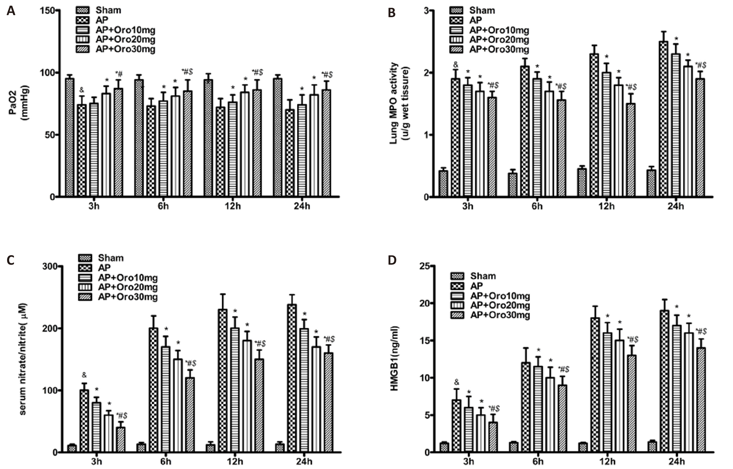
Figure 1. Preventive Administration of OroA Significantly Mitigated ALI in Pancreatitis Rats as Measured by PaO2 Levels, Nitrite/nitrate Concentrations, MPO Activity, and HMGB1 Expression. Data represent mean±SEM from 10 animals per group. Statistical comparisons were performed using one-way ANOVA followed by Tukey's post-hoc test (*P<0.05 vs. AP group; &P<0.01 vs. sham group; #P<0.05 vs. 10mg OroA-treated AP group; $P<0.05 vs. 20mg OroA-treated AP group).
3.2 BAL Fluid and NF-κB Activation
AP increased BAL protein concentration (1.2±0.1mg/mL vs. sham 0.4±0.05mg/mL, P<0.01) and neutrophil counts (2.5±0.3×10⁶cells/mL vs. sham 0.8±0.1×10⁶cells/mL, P<0.01). OroA treatment reduced both parameters, with the 30mg/kg dose achieving near-sham levels (P<0.05; Figure 2A and B). NF-κB activation in lung tissues was elevated in AP and suppressed by OroA (P<0.05; Figure 2D).
|
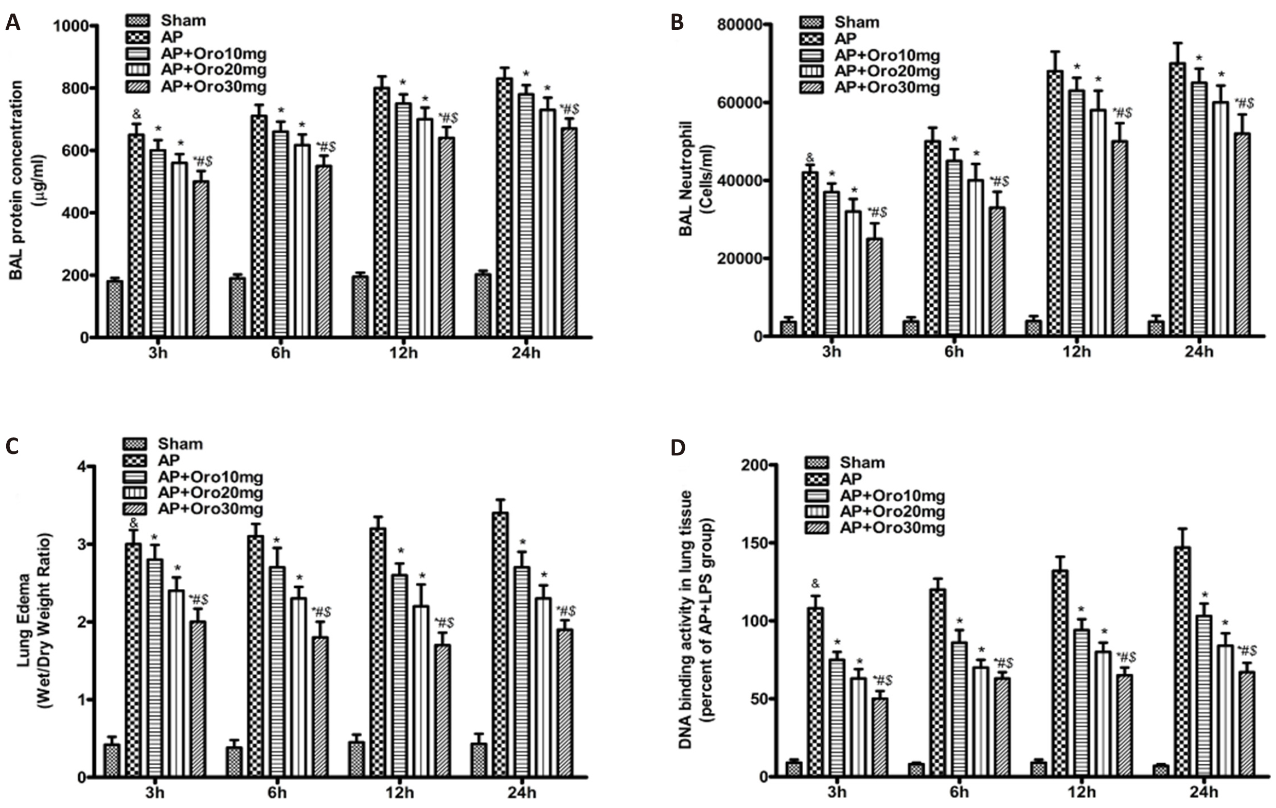
Figure 2. Prophylactic Treatment with OroA Modulated Inflammatory Responses in Pancreatitis-induced lung Injury, as Indicated by Reduced BAL Protein Concentration, Neutrophil Infiltration, Pulmonary Edema, and NF-κB Activation. All values are expressed as mean±SEM (n=10 per group). Significance was determined using one-way ANOVA with Tukey's multiple comparison test (&P<0.01 vs. sham; *P<0.05 vs. AP; #P<0.05 vs. 10mg OroA; $P<0.05 vs. 20mg OroA).
3.3 Lung Edema and Histopathology
AP induced significant pulmonary edema (W/D ratio: 4.2±0.3 vs. sham 2.8±0.2, P<0.01). OroA treatment dose-dependently reduced edema, with the 30 mg/kg group showing the lowest W/D ratio (3.1±0.2, P<0.05 vs. AP; Figure 2C). Histological analysis revealed alveolar damage, neutrophil infiltration, and septal thickening in AP lungs (Figure 3B). OroA treatment mitigated these changes in a dose-dependent manner (Figure 3C-E), with histological scores correlating with treatment efficacy (Figure 3F).
|
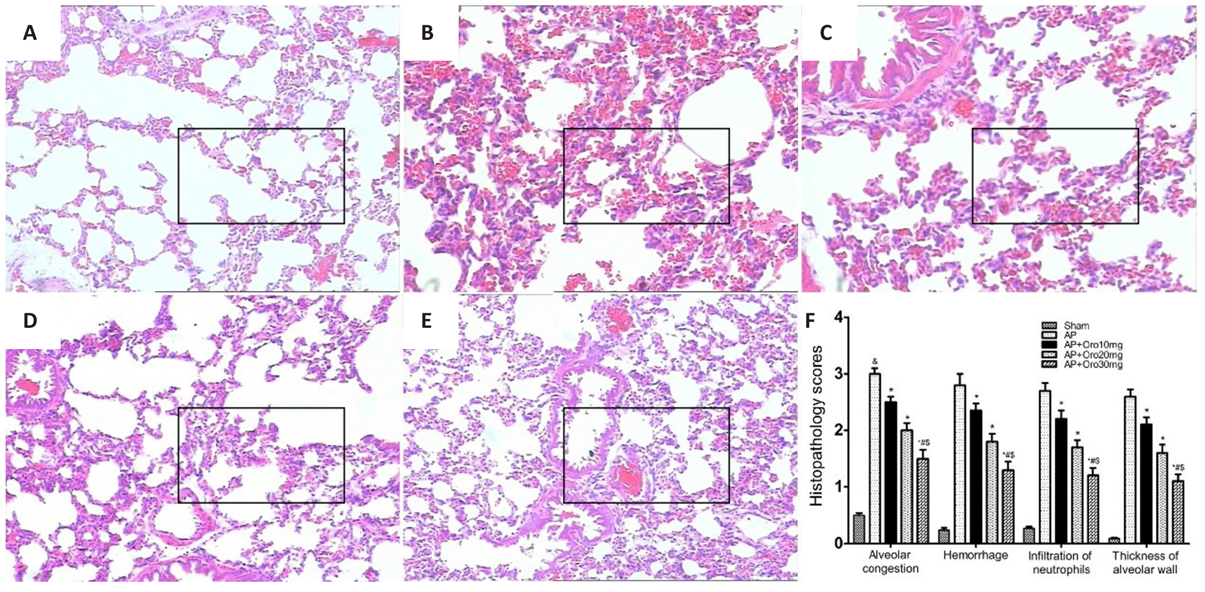
Figure 3. Histopathological Examination of Lung Tissues Using HE Staining. A: Sham-operated control group showing normal alveolar architecture. B: AP group demonstrating significant alveolar damage, neutrophil infiltration, and interstitial edema. C-E: AP rats treated with 10mg, 20mg, and 30mg OroA respectively, showing dose-dependent improvement in histological abnormalities. F: Semi-quantitative analysis of lung injury scores revealed significant reductions in the treatment groups. All data are presented as mean±SEM (n=10 per group). Statistical significance was determined by one-way ANOVA with Tukey's post-test (&P<0.01 vs. sham; *P<0.05 vs. AP; #P<0.05 vs. 10mg OroA; $P<0.05 vs. 20mg OroA). Original magnification: ×200.
4 DISCUSSION
AP-induced ALI remains a leading cause of mortality, with ALI/ARDS accounting for 30-40% of early deaths in severe AP cases[4,7]. This study provides novel evidence that OroA exerts protective effects against taurocholate-induced ALI through multi-targeted anti-inflammatory mechanisms. The observed reductions in PaO₂ decline, pulmonary edema, and histological damage suggest OroA may represent a promising therapeutic strategy for AP-related respiratory complications.
The protective effects of OroA were associated with suppression of the NF-κB signaling pathway, a central regulator of inflammatory gene expression. Inhibition of NF-κB translocation reduced downstream production of pro-inflammatory mediators including MPO, NO, and HMGB1[8]. These findings align with previous reports demonstrating OroA’s ability to block LPS-induced NF-κB activation in macrophage models. The observed dose-dependent reduction in HMGB1 levels is particularly significant, as this late-phase cytokine contributes to sustained inflammation and endothelial dysfunction in ALI[9-11].
Vascular endothelial injury, characterized by increased permeability and neutrophil infiltration, is a hallmark of ALI[12,13]. OroA treatment effectively mitigated taurocholate-induced endothelial dysfunction, as evidenced by reduced BAL protein concentration and neutrophil counts. This protective effect likely involves inhibition of iNOS-mediated NO overproduction, which disrupts endothelial integrity[14,15]. The suppression of NO metabolites (nitrite/nitrate) in treated animals supports this hypothesis, consistent with OroA’s previously reported anti-oxidative properties.
Histological analysis revealed that OroA ameliorated alveolar septal thickening, edema, and inflammatory cell infiltration in a dose-dependent manner. These morphological improvements correlated with functional outcomes, including restored PaO₂ levels and reduced lung W/D ratios. The observed histological score reductions (from 4.2±0.3 in AP to 3.1±0.2 in 30mg/kg OroA) suggest clinical translatability, as similar scoring systems are used in human ARDS grading[16,17].
While this study provides preclinical evidence for OroA’s efficacy, several limitations warrant further investigation. First, the optimal dosing regimen and long-term safety profile require validation in larger animal models. Second, mechanistic studies using gene knockout models or in vitro assays would clarify the precise role of NF-κB-independent pathways. Finally, translation to clinical settings necessitates evaluation of OroA’s pharmacokinetics and potential drug interactions.
Acknowledgements
This study was funded by Health Research Project 2022 (No. bzwj2022b003); and Bozhou Municipal Science and Technology Bureau 2024 Key R&D Project (No. bzzc2024025).
Conflicts of Interest
The authors declared no conflict of interest.
Ethics Statement
The experimental protocols were conducted in compliance with the guidelines of the National Institutes of Health and the United Kingdom Animals (Scientific Procedures) Act 1986. Ethical approval was obtained from the Institutional Animal Care and Use Committee of Anhui Medical University (Approval No. 202304).
Data Availability
All data generated or analyzed during this study are included in this published article.
Copyright Permissions
Copyright © 2025 The Author(s). Published by Innovation Forever Publishing Group Limited. This open-access article is licensed under a Creative Commons Attribution 4.0 International License (https://creativecommons.org/licenses/by/4.0), which permits unrestricted use, sharing, adaptation, distribution, and reproduction in any medium, provided the original work is properly cited.
Author Contribution
Wang H was responsible for designing the project, reviewing the literature, and processing the data; and Wang Z was responsible for writing and optimizing the manuscript.
Abbreviation List
ALI, Acute lung injury
AP, Acute pancreatitis
ARDS, Acute respiratory distress syndrome
BAL, Bronchoalveolar lavage
COX-2, Cyclooxygenase-2
HE, Hematoxylin-eosin
HMGB1, High mobility group box-1 protein
iNOS, Inducible nitric oxide synthase
MPO, Myeloperoxidase
NF-κB, Nuclear factor kappa B
NO, Nitric oxide
OroA, Oroxylin A
References
[1] Yu Q, Du Y, Li Z et al. Shenmai injection improves lung injury induced by severe acute pancreatitis in rats. Acad J Sec Mil Med Univ, 2013; 34: 1183-1186.[DOI]
[3] Shen L, Zhang L, Li H et al. Oroxylin A inhibits the generation of Tregs in non-small cell lung cancer. Oncotarget, 2017; 8: 49395-49408.[DOI]
[4] Li J, Tong D, Liu J et al. Oroxylin A attenuates cigarette smoke-induced lung inflammation by activating Nrf2. Int Immunopharmacol, 2016; 40: 524-529.[DOI]
[5] Lee A, Kang S, Park S et al. Anti-Allergic Effect of Oroxylin A from Oroxylum indicum Using in vivo and in vitro Experiments. Biomol Ther, 2016; 24: 283-90.[DOI]
[6] Zhou D, Diao B, Zhou W et al. Oroxylin A Inhibits Allergic Airway Inflammation in Ovalbumin (OVA)-Induced Asthma Murine Model. Inflammation, 2016; 39: 867-872.[DOI]
[7] Wei L, Yao Y, Zhao K et al. Oroxylin A inhibits invasion and migration through suppressing ERK/GSK-3beta signaling in snail-expressing non-small-cell lung cancer cells. Mol Carcinogen, 2016; 55: 2121-2134.[DOI]
[10] Yang X, Zhang F, Wang Y et al. Oroxylin A Inhibits Colitis-associated Carcinogenesis Through Modulating the IL-6/STAT3 Signaling Pathway. Inflamm Bowel Dis, 2013; 19: 1990-2000.[DOI]
[11] Zhang L, Chen Y, Wang L et al. Chloroquine relieves acute lung injury in rats with acute hemorrhagic necrotizing pancreatitis. J Huazhong Univ Sci Technolog Med Sci, 2013; 33: 357-360.[DOI]
[12] Yao J, Hu R, Sun J et al. Oroxylin A Prevents Inflammation-Related Tumor Through Down-Regulation of Inflammatory Gene Expression by Inhibiting NF-kappa B Signaling. Mol Carcinogen, 2014; 53: 145-158.[DOI]
[13] Hu H, Zhang Q, Kong B et al. Expression of pancreatic regenerating gene in lung and intestinal tissue correlates with the severity of disease in rats with acute necrotizing pancreatitis. Mol Med Rep, 2013; 7: 503-508.[DOI]
[14] Li H, Wu Y, Tseng T et al. Inhibition of miR-155 potentially protects against lipopolysaccharide-induced acute lung injury through the IRF2BP2-NFAT1 pathway. Am J Physiol Cell Physiol, 2020; 319: C1070-1081.[DOI]
[15] Zhi H, Jin X, Zhu H et al.Exploring the effective materials of flavonoids-enriched extract from Scutellaria baicalensis roots based on the metabolic activation in influenza A virus induced acute lung injury. J Pharmaceut Biomed, 2020; 177: 112876.[DOI]
[16] Zhi H, Zhu H, Zhang Y et al. In vivo effect of quantified flavonoids-enriched extract of Scutellaria baicalensis root on acute lung injury induced by influenza A virus. Phytomedicine, 2019; 57: 105-116.[DOI]
[17] Jin J, Chen S, Wang D et al. Oroxylin A suppresses influenza A virus replication correlating with neuraminidase inhibition and induction of IFNs. Biomed Pharmacother, 2018; 97: 385-394.[DOI]



 Copyright ©