
The Causal Association Between Blood Metabolites and Ovarian Cancer: Mendelian Randomization Study
Lin Zhu1#, Huimin Lu2#, Yanfeng Hou1, Yi Jin3,4#*, Xinghao Yu5,6*, Lijuan Wang1*
1Department of Obstetrics and Gynecology, The Fourth People’s Hospital of Changzhou, Jiangsu Province, China
2Department of outpatient and emergency, First Affiliated Hospital of Soochow University, Suzhou, Jiangsu Province, China
3The Wujin Clinical College of Xuzhou Medical University, Changzhou, Jiangsu Province, China
4Jiangsu Province Key Laboratory of Anesthesiology, Xuzhou Medical University, Xuzhou, Jiangsu Province, China
5National Clinical Research Center for Hematologic Diseases, Jiangsu Institute of Hematology, The First Affiliated Hospital of Soochow University, Suzhou, Jiangsu Province, China
6Center for Genetic Epidemiology and Genomics Research, Soochow UniversitySuzhou, Jiangsu Province, China
#These authors contributed equally to this work.
*Correspondence to: Lijuan Wang, PhD, Professor, Department of Obstetrics and Gynecology, The Fourth People’s Hospital of Changzhou City, Changzhou, 213000, Jiangsu Province, China; E-mail: wanglijuan_71@163.com
Xinghao Yu, PhD, Professor, National Clinical Research Center for Hematologic Diseases, Jiangsu Institute of Hematology, The First Affiliated Hospital of Soochow University, Soochow, 215006, Jiangsu Province, China;; E-mail: yuxinghao1994@suda.edu.cn
Yi Jin, PhD, Professor, The Wujin Clinical College of Xuzhou Medical University, Changzhou, 213000, Jiangsu Province, China; E-mail: jinyi@wjrmyy.cn
DOI: 10.53964/cme.2024013
Abstract
Objective: Ovarian cancer is closely related to human metabolism, but the causal effect of serum metabolites on its occurrence and development is still unknown.
Methods: Based on the publicly available Genome-Wide Association Studies (GWAS) abstract data set, this study used two-sample Mendelian randomization (MR) to determine the causal metabolites associated with ovarian cancer, and a comprehensive sensitivity analysis was used to verify the accuracy of the results. Finally, the metabolic pathway analysis of the causal metabolites that may affect the risk of ovarian cancer was carried out.
Results: Based on the summary level of the GWAS data set, the MR method was first used to identify 17 metabolites that are highly correlated with the risk of ovarian cancer, 16 of which are related to ovarian cancer subtypes. Further sensitivity analysis excluded the influence of heterogeneity and horizontal pleiotropy, and confirmed the causal effects of 9 metabolites. Metabolic pathway analysis shows that tryptophan metabolic pathway may play a key role in invasive epithelial ovarian cancer, alanine, aspartic acid and glutamate metabolism, citrate cycle, glyoxylic acid and dicarboxylate Metabolism is closely related to mucinous ovarian cancer, and caffeine metabolism can affect the occurrence and development of low-grade potential ovarian cancer.
Conclusion: This study comprehensively assessed the influence of blood metabolites on the risk of ovarian cancer and its different subtypes through genetic methods.
Keywords: metabolites, ovarian cancer, Mendelian randomization, metabolic pathways
1 INTRODUCTION
Ovarian cancer (OC) is the second most common cause of death among gynecologic cancers worldwide[1]. Common symptoms of OC encompass bloating, pelvic pain, abdominal swelling, and diminished appetite. The prognosis for OC is predominantly unfavorable, largely because its early stages often go undetected due to the absence of pronounced early symptoms and the lack of established screening tests. The precise cause of OC remains elusive, with potential risk factors being the timing of ovulation, age, hormonal influences, and genetic predispositions[2]. Of these, epithelial OC accounts for 85%-90% of OCs, and only few risk factors associated with epithelial OC have been identified in previous observational epidemiologic studies, and most of the previous studies did not perform subgroup analyses[3]. Subgroup analysis is essential in clinically diverse histotypes, and previous analyses have reported heterogeneity in the association of risk factors with subgroups[4-6]. Furthermore, it is unclear whether the reported risk factors have a causal effect on OC, given that traditional observational designs are susceptible to residual confounding and reverse causation[7].
Metabolites play a very important role in cellular functions as products and substrates in metabolic pathways, including cell proliferation and apoptosis, which are closely related to cancer development and progression. Previous studies have performed a series of metabolomic studies on OC using different techniques. One study that analyzed serum from OC patients identified pentyl glucuronide as a potential biomarker for OC[8]. Another study utilizing H nuclear magnetic resonance spectroscopy showed significantly elevated levels of metabolites such as acetoacetate, acetone and 3-hydroxybutyrate in OC patients[9]. Traditional clinical studies have difficulty establishing a causal relationship between blood metabolites and OC due to unavoidable confounding factors. In recent years, numerous studies have combined metabolomics with high-throughput genotyping to estimate the effects of genetic variants on metabolic phenotypes through Genome-Wide Association Studies (GWAS) and identified thousands of genetic loci associated with metabolic phenotypes[10,11]. Recently, Mendelian randomization (MR) based on large GWAS datasets has proven to be a powerful tool for assessing the etiology of complex diseases, as they can effectively control for unknown confounders[12,13]. There are still no studies that comprehensively assess the causal role of metabolic profiling on OC risk.
In this paper, we systematically assessed the causal relationship between blood metabolites and invasive epithelial OC (IEOC) in conjunction with large-scale summary statistics from previous GWAS[14], and explored common metabolic mechanisms among different OC types. Specifically, the study performed a comprehensive two-sample MR analysis to explore the causative metabolites of OC and its different subtypes. The biological functions of the identified pathogenic metabolites were further determined by metabolic pathway analysis.
2 MATERIALS AND METHODS
2.1 Data
In this study, we utilized the most comprehensive GWAS summary statistics on human blood metabolite profiles, obtained from the Metabolomics GWAS server (http://metabolomics.helmholtz-muenchen.de/gwas/)[10]. After stringent quality control, the dataset comprised association analyses for 486 metabolites (309 known) based on SNP data from 7,824 European individuals. Since the biological functions of the unknown metabolites are not yet clear, we focused on the causal relationships between the 309 known metabolites and OC. These metabolites are categorized into 60 subclasses and 8 major groups, as defined by the KEGG pathway. GWAS summary statistics, including effect size, standard error, and sample size, are available for approximately 2.1 million SNPs.
We obtained GWAS summary statistics for OC from the OC Association Consortium (OCAC), encompassing 25,509 women with IEOC and 40,941 controls of European ancestry[15]. The dataset includes 22,406 IEOC cases and covers histologic subtypes such as high-grade serous carcinoma (n=13,037), low-grade serous carcinoma (n=1,012), mucinous carcinoma (n=1,417), endometrioid carcinoma (n=2,810), and clear cell carcinoma (n=1,366). Additionally, 3,103 cases of potentially malignant low-grade carcinomas were analyzed, comprising 1,954 serous and 1,149 mucinous carcinomas. All OCAC studies had ethical approval, and participants provided written informed consent. Data sources and additional details are outlined in Table 1.
Table 1. Detailed information of GWAS summary data
Traits |
Year |
Sample Size |
SNPs |
PMID |
|
Case/Control |
|||||
Blood Metabolites |
2014 |
7,824 |
2,545,661 |
24816252 |
|
Invasive epithelial ovarian cancer (IEOC) |
2017 |
22,406/40,941 |
18,549,275 |
28346442 |
|
|
High grade plasma (HGS) |
13,307/40,941 |
18,549,275 |
|
|
|
Low grade plasma (LGS) |
1,012/40,941 |
18,549,275 |
|
|
|
Mucinous (MS) |
1,417/40,941 |
18,549,275 |
|
|
|
Endometrial |
2,810/40,941 |
18,549,275 |
|
|
|
Clear cell (CC) |
1,366/40,941 |
18,549,275 |
|
|
|
Low potential malignancy (LMP) |
3,103/40,941 |
18,549,275 |
|
|
2.2 Selection of Instrumental Variables
Consistent with previous studies, this paper utilized the clumping program of PLINK software and selected instrumental variables for each metabolite with a loose significance threshold[16,17]. Specifically, the clump function used the 1,000 Genomes Project as the reference dataset[18], the significance threshold was set to 10-5, the chain disequilibrium r2 threshold was 0.1, and the window was set to 500KB, consistent with previous studies[19,20]. A total of 3 to 631 independent SNPs were selected as instrumental variables for 309 metabolites, with one metabolite having no significant loci. For the reverse MR analysis on OC, instrumental variable selection followed a similar process, with a significance threshold of 5×10⁻⁸. To ensure SNP strength, we calculated the proportion of phenotypic variance explained (PVE) and F-statistics, excluding those with F-statistics below 10. Additionally, SNPs strongly associated with outcomes (P<0.05 after correction), with abnormal effect sizes, or in MHC regions were also removed.
2.3 Statistical Analysis
To assess the causal effect of potentially pathogenic metabolites on OC, we conducted a two-sample MR analysis. Heterogeneity was evaluated using Cochran’s Q statistic, with random-effects IVW applied when the null hypothesis was rejected, and fixed-effects otherwise[21]. Given multiple comparisons, metabolites with P<0.007 (0.05/7) were deemed causal for OC, while those between 0.05 and 0.007 indicated potential causality. Sensitivity analyses-including the weighted median method[22], maximum likelihood method[23], and MR-Egger regression[24] were used to control for horizontal pleiotropy. Additionally, LOO analysis and MR-PRESSO were performed to detect outliers due to horizontal pleiotropy[25]. To exclude potential bidirectional associations, reverse MR analysis was also conducted to estimate OC’s causal impact on the identified metabolites.
To explore the functions and pathways of the identified metabolites, MetaboAnalyst 4.0 (https://www.metaboanalyst.ca/)[26] was used in this paper to perform metabolic pathway analysis of 18 metabolites causally associated with IEOC and to reveal the possible pathways of metabolites enriched for different subtypes of IEOC, respectively. The pathway analysis tool uses high-quality KEGG[27] and Small Molecule Pathway Database (SMPDB) metabolic pathways[28] as the back-end knowledge base and integrates powerful pathway enrichment analysis and pathway topology analysis. The significance level of metabolic pathways was set at 0.05.
Statistical analyses were performed in R 3.5.3 software, MR analysis was performed using the MR package[29] and MR-PRESSO was performed using the MR-PRESSO package[30].
3 RESULTS
3.1 Causal Effects of Potentially Pathogenic Metabolites on OC and its Subtypes were Examined by MR analysis.
To explore the causal effects of metabolites on OC, we assessed the causal effects among 486 metabolites using the IVW method (Figure 1). The instrumental variables selected all had F-statistics greater than 10, which can be considered as strong instrumental variables. The results revealed that 18 metabolites were associated with IEOC (P<0.05). Among them, seven metabolites belonged to the lipid pathway, eight to the amino acid pathway, one to the peptide pathway, one to the xenobiotic pathway and one to the carbohydrate pathway. Under a strict significance level (P<0.05/7=0.007), 2 metabolites remained significant, C-glycosyltryptophan (risk ratio (OR)=3.510, P=0.002) and indoleacetic acid (OR=0.610, P=0.005). In the results of OC subtypes, C-glycosyltryptophan was found to be a potential risk factor for highly plasma (OR=2.593, P=0.047), mucinous (OR=18.107, P=0.017), and low-grade potentially malignant (OR=12.102, P=0.005) cancers. There was a negative causal relationship between indoleacetic acid and highly plasma carcinoma (OR=0.600, P=0.014) as well as clear cell carcinoma (OR=0.312, P=0.032). Results for different OC subtypes showed that 15 relevant causal metabolites were identified after Bonferroni (P<0.007) correction. There were causal associations between 2 of the metabolites and low-grade plasmacytoid carcinoma, where mannitol was a protective factor for low-grade plasmacytoid carcinoma (OR=0.122, P=6.85×10⁻⁴) while bile acids were a risk factor (OR=2.132, P=0.004). For causality with hyperplasia, three metabolites are causally involved (1-methylxanthine, nonanoyl carnitine, and palmitoleate (16:1n7, respectively), where 1-methylxanthine was a protective factor for highly plasma carcinoma (OR=0.647, P=0.003) while nonanoyl carnitine and palmitoleate (16:1n7) were risk factors (ORs of 1.34, P=0.003 and 1.969, P=0.005). Mucinous OC results showed that glutamyl tyrosine reduced the risk of OC (OR=0.151, P=0.004), while 4-acetylaminophenol sulphate (OR=1.079, P=0.005) as well as ADpSGEGDFXAEGGGVR (OR=3.705, P=0.005) increased its risk. Mannose, palmitoyl carnitine and 1-methyluronate were all protective factors for low-grade malignant potential OC with ORs of 0.28 (P=5.58E-4), 0.221 (P=0.004) and 0.521 (P=0.005), respectively. Finally, five causal metabolites were causally associated with endometrioid OC, with arachidonic acid (20:4n6) (OR=0.238, P=2.93×10⁻⁴) and inosine (OR=0.647, P=0.007) as protective factors, while 3-(3-hydroxyphenyl) propionate (OR=1.442, P=0.004), 1,5-anhydroglucosol (1,5-AG) (OR=2.442, P=0.004), and C-glycosyltryptophan (OR=12.102, P=0.005) were risk factors.
|
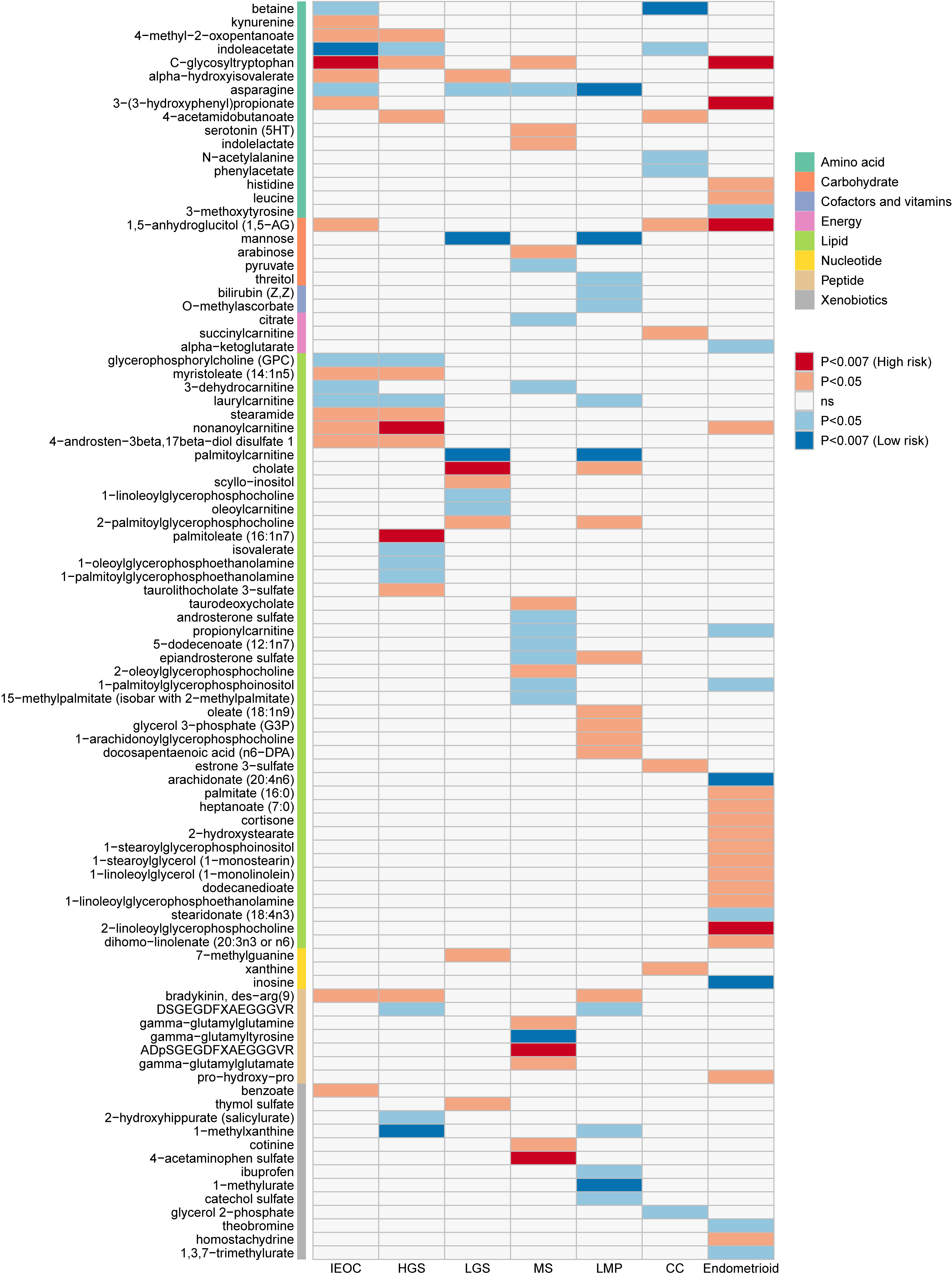
Figure 1. Causal Effects of Metabolites with Ovarian Cancer and its Subtypes. IEOC: invasive epithelial ovarian cancer; HGS, High grade plasma; LGS, Low grade plasma; MS, Mucinous; CC, Clear cell; LMP, Low potential malignancy.
In this paper, multiple sensitivity analyses were further performed on the identified causal associations (Table 2), which showed that the sensitivity analyses for most of the metabolites were in agreement with the IVW results.The intercept term of the MR-Egger test showed horizontal pleiotropy for the associations of two metabolites with OC: C-glycosyltryptophan with IEOC, and 1-methylxanthine with high plasmaticity, respectively. The association between C-glycosyltryptophan and IEOC was further assessed using MR-PRESSO, and the results were consistent with the IVW results (OR=3.510, 95% CI=1.510-8.155, P=0.010). One instrumental variable had a strong correlation with outcome (Z=-2.574, P=0.010), and horizontal pleiotropy disappeared when this SNP was excluded (P intercept=0.074). The association between 1-methylxanthine and high plasmaticity was similarly assessed using MR-PRESSO (OR=0.647, 95% CI=0.472 to 0.887, P=0.017). One instrumental variable was strongly associated with outcome (Z=-2.768, P=0.006), and horizontal pleiotropy remained after excluding this SNP (P intercept=0.020). Further results of weighted median analysis revealed that nine metabolites were still causally associated with OC and its subtypes and were not affected by horizontal pleiotropy, so they could be considered as the final causal metabolites. Finally, reverse MR analysis ruled out the possibility of a causal effect of OC on the metabolites.
Table 2. Sensitivity Analysis of Association of Causal Metabolites with Ovarian Cancer (PIVW < 0.007)
Traits |
Metabolites |
SNP |
heterogeneity Test P |
IVW |
Weighted Median |
MR-Egger |
|||||
OR (95%CI) |
P |
OR (95%CI) |
P |
OR (95%CI) |
P |
Intercept |
P |
||||
IEOC |
C-glycosyltryptophan |
18 |
0.315 |
3.51 (1.589, 7.754) |
0.002 |
5.724 (1.644, 19.927) |
0.006 |
0.669 (0.126, 3.554) |
0.637 |
0.022 |
0.033 |
IEOC |
Indoleacetic acid ester |
22 |
0.112 |
0.61 (0.433, 0.86) |
0.005 |
0.652 (0.394, 1.076) |
0.094 |
0.645 (0.311, 1.338) |
0.253 |
-0.001 |
0.857 |
Endometrial |
C-Glycosyltryptophan |
18 |
0.393 |
12.102 (2.162, 67.727) |
0.005 |
4.318 (0.304, 61.261) |
0.280 |
1.441 (0.038, 54.866) |
0.846 |
0.029 |
0.211 |
HGS |
Nonyl carnitine |
22 |
0.148 |
1.34 (1.103, 1.627) |
0.003 |
1.488 (1.067, 2.074) |
0.019 |
1.318 (0.843, 2.061) |
0.240 |
0.001 |
0.934 |
Endometrial |
3-(3-Hydroxyphenyl)propionate |
11 |
0.849 |
1.442 (1.126, 1.847) |
0.004 |
1.581 (1.069, 2.338) |
0.022 |
1.384 (0.866, 2.211) |
0.174 |
0.004 |
0.838 |
CC |
Betaine |
24 |
0.619 |
0.175 (0.049, 0.616) |
0.007 |
0.078 (0.011, 0.551) |
0.011 |
0.215 (0.01, 4.746) |
0.330 |
-0.004 |
0.885 |
Endometrial |
1,5-Anhydroglucitol (1,5-AG) |
41 |
0.837 |
2.442 (1.328, 4.491) |
0.004 |
1.752 (0.701, 4.379) |
0.230 |
1.541 (0.347, 6.852) |
0.570 |
0.009 |
0.508 |
HGS |
Palmitoleate (16:1n7) |
10 |
0.798 |
1.969 (1.228, 3.159) |
0.005 |
1.778 (0.945, 3.347) |
0.074 |
1.472 (0.51, 4.251) |
0.475 |
0.008 |
0.548 |
Endometriotic |
Arachidonic acid (20:4n6) |
26 |
0.219 |
0.238 (0.109, 0.517) |
0.000 |
0.81 (0.211, 3.112) |
0.758 |
0.109 (0.025, 0.468) |
0.006 |
0.017 |
0.210 |
Endometriotic |
Inosine |
10 |
0.493 |
0.647 (0.473, 0.886) |
0.007 |
0.582 (0.383, 0.883) |
0.011 |
0.497 (0.176, 1.41) |
0.225 |
0.021 |
0.617 |
MS |
Gamma-glutamyl tyrosine |
45 |
0.478 |
0.151 (0.042, 0.547) |
0.004 |
0.136 (0.02, 0.918) |
0.041 |
0.016 (0, 0.984) |
0.054 |
0.024 |
0.266 |
LMP |
Cholate |
9 |
0.212 |
2.132 (1.279, 3.556) |
0.004 |
2.183 (1.011, 4.712) |
0.047 |
1.615 (0.356, 7.329) |
0.554 |
0.022 |
0.704 |
LMP |
Mannose |
26 |
0.084 |
0.122 (0.036, 0.41) |
0.001 |
0.08 (0.01, 0.626) |
0.016 |
0.037 (0.002, 0.757) |
0.043 |
0.029 |
0.384 |
LMP |
Mannose |
26 |
0.420 |
0.28 (0.136, 0.577) |
0.001 |
0.338 (0.103, 1.109) |
0.073 |
0.396 (0.085, 1.848) |
0.250 |
-0.008 |
0.622 |
HGS |
1-Methylxanthine |
14 |
0.254 |
0.647 (0.486, 0.861) |
0.003 |
0.657 (0.378, 1.14) |
0.135 |
0.383 (0.226, 0.651) |
0.002 |
0.021 |
0.031 |
LMP |
1-Methyluric acid ester |
14 |
0.169 |
0.521 (0.329, 0.825) |
0.005 |
0.596 (0.264, 1.347) |
0.214 |
0.45 (0.187, 1.086) |
0.099 |
0.008 |
0.684 |
LMP |
Palmitoyl carnitine |
9 |
0.713 |
0.221 (0.08, 0.61) |
0.004 |
0.24 (0.061, 0.95) |
0.042 |
0.368 (0.041, 3.333) |
0.374 |
-0.013 |
0.609 |
MS |
4-Acetaminophen Sulfate |
23 |
0.728 |
1.079 (1.024, 1.138) |
0.005 |
1.066 (0.988, 1.151) |
0.097 |
1.036 (0.899, 1.192) |
0.627 |
0.021 |
0.535 |
MS |
ADpSGEGDFXAEGGGVR |
7 |
0.150 |
3.705 (1.49, 9.216) |
0.005 |
2.894 (0.766, 10.928) |
0.117 |
0.221 (0.013, 3.662) |
0.333 |
0.118 |
0.082 |
3.2 Metabolic Pathway Analysis
In order to explore the relationship between metabolites and OC in a comprehensive manner, this study further performed metabolic pathway analysis of the causal metabolites identified by MR for OC and its subtypes (Table 3). For IEOC, we detected the tryptophan metabolic pathway belonging to both KEGG and SMPDB (P=0.038). Three significant pathways were found in mucinous OC: alanine, aspartate and glutamate metabolism (P=0.001), citrate cycle (TCA cycle) (P=0.010), and glyoxylate and dicarboxylate metabolism (P=0.024). Finally, an important role of caffeine metabolic pathway was detected in low malignant potential (P=0.002).
Table 3. Metabolic Pathway Analysis of Causal Metabolites
Subgroups |
Pathway Name |
P |
Impact |
Details |
IEOC |
Tryptophan metabolism |
0.038 |
0.094 |
KEGG SMP |
MS |
Alanine, aspartate and glutamate metabolism |
0.001 |
0.000 |
KEGG SMP SMP SMP |
MS |
Citrate cycle (TCA cycle) |
0.010 |
0.137 |
KEGG SMP |
MS |
Glyoxylate and dicarboxylate metabolism |
0.024 |
0.032 |
KEGG |
LMP |
Caffeine metabolism |
0.002 |
0.308 |
KEGG SMP |
3.3 Validation of Identified Metabolites
We extracted data from another metabolite GWAS to verify the identified metabolites, and the results are shown in Figure 2. The results showed that C-Glycosyltryptophan was consistent with the discovery set, and elevated metabolite levels increased the risk of IEOC (OR=1.064, P=0.016). Although no proteFctive effect of Mannose was found on LGS and LMP, the validation set found that it had a protective effect on MS (OR = 0.814, P=0.049). The significant causal association between Cholate and LMP, inosine and Endometrioid, and 1-methylxanthine and HGS was also confirmed (P<0.05); the association between 4-Acetaminophen Sulfate and MS was not found in the validation set (P>0.05).
|
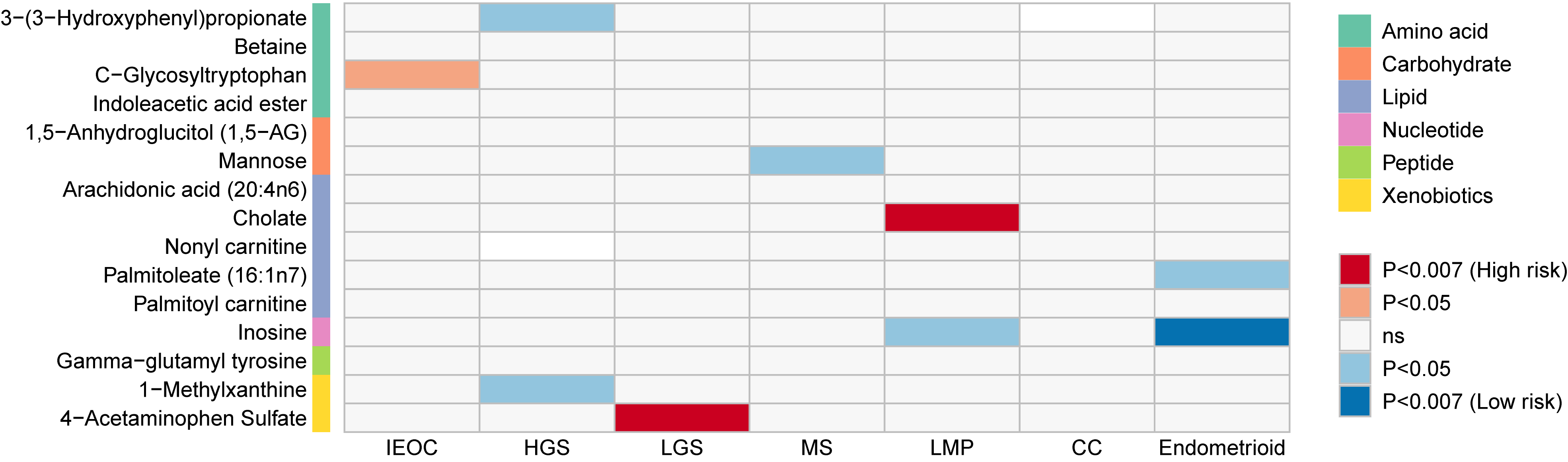
Figure 2. Validation of Causal Associations Between Identified Metabolites with Ovarian Cancer and its Subtypes.
4 DISCUSSION
To date, this is the first study to integrate large-scale GWAS summary datasets to systematically reveal the mechanisms of metabolites on OC development from a genetic perspective. This study provides strong evidence that blood metabolites can influence OC risk through an integrated genetic approach based on large GWAS pooled data. Utilizing SNPs as instrumental variables, the integration of multiple two-sample MR methods demonstrated nine causal metabolites associated with OC risk, including C-glycosyltryptophan, nonanoyl carnitine, 3-(3-hydroxyphenyl) propionate, betaine, inosine, γ-glutamyl tyrosine, cholate, mannose, and palmitoyl carnitine. A variety of metabolites may be potentially associated with OC development. KEGG and SMPDB pathway analyses demonstrated that metabolic pathways such as caffeine metabolism, biosynthesis of valine, leucine, and isoleucine, aminoacyl-tRNA biosynthesis, and tryptophan metabolism may play key roles in the metabolism of OC or its subtypes. In addition, the association between 1-methylxanthine and high plasmaticity may be affected by horizontal pleiotropy and therefore was not considered in this study at this time.
This finding underscores the potential of integrating metabolomic biomarkers into existing clinical tools, which could enable more precise risk stratification and personalized treatment planning[31]. This finding underscores the potential of integrating metabolomic biomarkers into existing clinical tools, which could enable more precise risk stratification and personalized treatment planning. The identification of pseudouridine and triglycerides as novel risk factors for OC offers further insights into disease mechanisms[32]. The association between elevated pseudouridine levels and high-grade serous tumors highlights its potential as a biomarker for identifying aggressive OC types. Clinically, pseudouridine’s role in translational fidelity and its presence in tumor-specific splicing could inform targeted therapeutic strategies, particularly in the management of rapidly progressing OC[32]. An MR study identified 31 metabolites with significant causal effects on OC, including Androsterone sulfate and Propionylcarnitine, which promote OC, and X-12,093 and Octanoylcarnitine, which are protective. These findings suggest potential biomarkers for clinical validation, though OC subtypes were not differentiated[33], which did not consider differences between different OC subtypes.
Studies have shown that abnormalities in glycosylation are involved in the pathophysiology of malignant tumors, and compounds that are glycosylated are used as potential biomarkers for the early detection of disease, as well as for assessing the efficacy of therapies for cancer, diabetes, and other diseases[34]. Our study’s findings align with this body of work, particularly the involvement of C-glycosyltryptophan in the tryptophan metabolic pathway. Given its connection to immune evasion via indoleamine 2,3-dioxygenase, targeting this pathway could bolster immune response and offer new therapeutic avenues[35]. These metabolites, especially those linked with immune modulation, provide promising targets for therapeutic interventions aimed at reactivating immune responses against OC cells. Regarding methylxanthines, our discovery of their protective effect against OC aligns with the observed anticancer properties of coffee and other sources rich in antioxidants[36]. This suggests that dietary interventions involving methylxanthine-rich foods could serve as supplementary preventive strategies for OC, a concept supported by existing evidence associating decaffeinated coffee with reduced OC risk[37]. Despite no significant association between nonylcarnitine and high-risk OC in our study, the documented role of carnitine metabolism in OC progression warrants further exploration. Given carnitine palmitoyltransferase’s role in fatty acid metabolism, targeting this pathway could impede OC development, particularly in high-grade cases where metabolic reprogramming is pronounced[38,39]. Furthermore, existing studies suggest that targeting palmitoylcarnitine could leverage its ability to induce oxidative stress in cancer cells, providing a possible adjunctive treatment avenue[40,41]. Finally, the findings regarding mannose highlight its therapeutic potential. As an oral supplement, mannose’s ability to modulate the AKT and ERK1/2 pathways could be leveraged to slow OC progression, especially in combination with standard therapies[42]. Clinically, mannose supplementation could be explored as an adjunctive strategy to enhance the efficacy of conventional treatments.
Protective metabolites like arachidonic acid and inosine suggest pathways for resisting oncogenesis. Arachidonic acid's role in inflammation and cell signaling hints at using inflammation modulation as a preventive strategy for OC[43], despite past findings that AA and its metabolites promote cancer[44-46]. Similarly, inosine’s immunomodulatory properties open avenues for enhancing immune targeting of neoplastic cells[47]. Conversely, metabolites such as 3-(3-Hydroxyphenyl) propionate, 1,5-anhydroglucosol, and C-glycosyltryptophan emerge as risk factors, highlighting metabolic vulnerabilities to cancer. The association of 3-(3-Hydroxyphenyl) propionate with cancer risk may point to gut microbiota's role in oncogenesis, while 1,5-AG links metabolic health to OC, supporting metabolic interventions as prevention. Although specific links between these metabolites and OC are not well-studied, 1,5-AG has been associated with cancer mortality risks related to glucose levels[48]. The presence of C-glycosyltryptophan in OC patients suggests an amino acid metabolism role in cancer progression[49]. This study using UPLC to measure plasma C-glycosyltryptophan found that plasma CMW was significantly higher in patients with malignant or borderline OC than in the benign tumor group and normal controls[49].
Notably, there were causal associations between some metabolites and multiple OC subtypes, e.g., indoleacetate was a protective factor for IEOC, HGS and CC subtypes; and the metabolite C-Glycosyltryptophan was a risk factor for IEOC, HGS, LGS and Endometriotic. These results provide some evidence for OC prevention and treatment.
There exist limitations in our research. This study needs further clinical trials to confirm the new findings in the study, e.g., there is no report of nonanoyl carnitine associated with cancer metabolism so far. Further validation by biofunctional assays is also needed, which is beyond the scope of this study. In addition, due to the effective sample size of the metabolite database, the use of a more relaxed significance threshold in this study aimed to identify more metabolites that may be related to the mechanism of OC development and to fully elucidate the pathway of metabolite action on OC. Due to the lack of individual data, further stratification of the population (e.g., age, BMI, etc.) was not possible to investigate the effect of metabolite levels on OC risk in different populations. After searching the GWAS catalog and PubMed, the current GWAS of subtype data is very limited, and we cannot obtain metabolomic studies based on individual data. The OC data we currently use is still the largest GWAS study with the largest sample size. Therefore, we cannot perform a valid verification here. Since no significant causal metabolites were found after multiple correction, we chose a slightly stricter significance level (P<0.05/7) to report the results and verified the stability of the results based on multiple sensitivity analyses. However, it is worth noting that using a loose significance level will lead to certain false positives. In addition, due to the limited statistical power of GWAS at the metabolite level, we chose a more common threshold for instrumental variable screening, which may also increase false positives. Therefore, other studies need to be more cautious when drawing conclusions.
In summary, alterations in the levels of metabolites such as lipid metabolism and amino acid metabolism play an important role in the development of OC. This study explores causal metabolites of OC and its subtypes at the gene level based on a large publicly available GWAS public dataset, which will provide a new perspective for further OC etiology studies and help people to make early interventions to reduce the risk of OC.
Acknowledgements
Jin Y was supported by Open Funding Project of Jiangsu Provincial Key Laboratory of Anesthesiology, Xuzhou Medical University (XZSYSKF2023011); The Changzhou Sci&Tech Program (No. CJ20242012). And thank all the studies for making the summary association statistics data publicly available.
Author Contribution
Wang L and Yu X designed the study. Zhu L obtained the data; Zhu L and Lu H cleared up the datasets; Yu X, Zhu L and Hou Y mainly performed the data analyses; Wang L, Yu X, Zhu L, Lu H and Hou Y drafted the manuscript, and all authors read and approved the final manuscript.
Conflicts of Interest
All authors declared no potential conflicts of interest.
Data Sharing Statement
Data are available in public, open access repositories corresponding to the original studies (e.g., GWAS catalog).
Abbreviation List
AA, Arachidonic acid
CC, Clear cell (carcinoma)
CI, Confidence interval
GWAS, Genome-Wide Association Studies
HGS, High grade serous (carcinoma)
IEOC, Invasive epithelial ovarian cancer
IVW, Inverse variance weighted
KEGG, Kyoto encyclopedia of genes and genomes
LGS, Low grade serous (carcinoma)
LMP, Low malignant potential
LOO, Leave
MHC, Major histocompatibility complex
MR, Mendelian randomization
MS, Mucinous (carcinoma)
NMR, Nuclear magnetic resonance
OC, Ovarian cancer
OCAC, Ovarian cancer association consortium
OR, Odds ratio
PVE, Proportion of phenotypic variance explained
SMPDB, Small molecule pathway database
SNP, Single nucleotide polymorphism
TCA, Tricarboxylic acid (cycle)
UPLC, Ultra performance liquid chromatography
References
[1] Lheureux S, Braunstein M, Oza AM. Epithelial ovarian cancer: Evolution of management in the era of precision medicine. CA-Cancer J Clin, 2019; 69: 280-304.[DOI]
[2] Karnezis AN, Cho KR, Gilks CB et al. The disparate origins of ovarian cancers: pathogenesis and prevention strategies. Nat Rev Cancer, 2017; 17: 65-74.[DOI]
[3] Lheureux S, Gourley C, Vergote I et al. Epithelial ovarian cancer. Lancet, 2019; 393: 1240-1253.[DOI]
[4] Corvigno S, Badal S, Spradlin ML et al. In situ profiling reveals metabolic alterations in the tumor microenvironment of ovarian cancer after chemotherapy. NPJ Precis Oncol, 2023; 7: 115.[DOI]
[5] Wang X, Xie W, Zhao D et al. Molecular Subtypes of Ovarian Cancer Based on Lipid Metabolism and Glycolysis Reveals Potential Therapeutic Targets. Fronti biosci-Landmrk, 2023; 28: 253.[DOI]
[6] Murali R, Balasubramaniam V, Srinivas S et al. Deregulated Metabolic Pathways in Ovarian Cancer: Cause and Consequence. Metabolites, 2023; 13: 560.[DOI]
[7] Kyriacou DN, Lewis RJ. Confounding by Indication in Clinical Research. Jama, 2016; 316: 1818-1819.[DOI]
[8] Chen J, Zhang X, Cao R et al. Serum 27-nor-5β-cholestane-3,7,12,24,25 pentol glucuronide discovered by metabolomics as potential diagnostic biomarker for epithelium ovarian cancer. J Proteome Res, 2011; 10: 2625-2632.[DOI]
[9] Garcia E, Andrews C, Hua J et al. Diagnosis of early stage ovarian cancer by 1H NMR metabonomics of serum explored by use of a microflow NMR probe. J Proteome Res, 2011; 10: 1765-71.[DOI]
[10] Shin SY, Fauman EB, Petersen AK et al. An atlas of genetic influences on human blood metabolites. Nat Genet, 2014; 46: 543-50.[DOI]
[11] Schlosser P, Li Y, Sekula P et al. Genetic studies of urinary metabolites illuminate mechanisms of detoxification and excretion in humans. Nat Genet, 2020; 52: 167-176.[DOI]
[12] Elliott J, Bodinier B, Bond TA et al. Predictive Accuracy of a Polygenic Risk Score-Enhanced Prediction Model vs a Clinical Risk Score for Coronary Artery Disease. Jama, 2020; 323: 636-645.[DOI]
[13] Xia J, Xie SY, Liu KQ et al. Systemic evaluation of the relationship between psoriasis, psoriatic arthritis and osteoporosis: observational and Mendelian randomisation study. Ann Rheum Dis, 2020, 79: 1460-1467.[DOI]
[14] Sudlow C, Gallacher J, Allen N et al. UK biobank: an open access resource for identifying the causes of a wide range of complex diseases of middle and old age. PLoS Med, 2015; 12: e1001779.[DOI]
[15] Phelan CM, Kuchenbaecker KB, Tyrer JP et al. Identification of 12 new susceptibility loci for different histotypes of epithelial ovarian cancer. Nat Genet, 2017; 49: 680-91.[DOI]
[16] Yang J, Yan B, Zhao B et al. Assessing the causal effects of human serum metabolites on 5 major psychiatric disorders. Schizophrenia Bull, 2020, 46: 804-813.[DOI]
[17] Purcell S, Neale B, Todd-Brown K et al. PLINK: a tool set for whole-genome association and population-based linkage analyses. Am J Hum Genet, 2007; 81: 559-75.[DOI]
[18] Consortium GP. A global reference for human genetic variation. Nature, 2015; 526: 68.[DOI]
[19] Yu XH, Cao RR, Yang YQ, Lei SF. Identification of causal metabolites related to multiple autoimmune diseases. Hum Mol Genet, 2022; 31: 604-13.[DOI]
[20] Yang J, Yan B, Zhao B et al. Assessing the causal effects of human serum metabolites on 5 major psychiatric disorders. Schizophrenia Bull, 2020; 46: 804-813.[DOI]
[21] Burgess S, Small DS, Thompson SG. A review of instrumental variable estimators for Mendelian randomization. Stat Methods Med Res, 2017; 26: 2333-2355.[DOI]
[22] Bowden J, Smith GD, Haycock PC et al. Consistent Estimation in Mendelian Randomization with Some Invalid Instruments Using a Weighted Median Estimator. Genet Epidemiol, 2016; 40: 304-314.[DOI]
[23] Burgess S, Butterworth A, Thompson SG. Mendelian Randomization Analysis With Multiple Genetic Variants Using Summarized Data. Genet Epidemiol, 2013; 37: 658-65.[DOI]
[24] Burgess S, Thompson SG. Interpreting findings from Mendelian randomization using the MR-Egger method. Eur J Epidemiol, 2017; 32: 377-389.[DOI]
[25] Verbanck M, Chen C-Y, Neale B et al. Detection of widespread horizontal pleiotropy in causal relationships inferred from Mendelian randomization between complex traits and diseases. Nat Genet, 2018; 50: 693-698.[DOI]
[26] Chong J, Soufan O, Li C, Caraus I, Li S, Bourque G, et al. MetaboAnalyst 4.0: towards more transparent and integrative metabolomics analysis. Nucleic Acids Res, 2018; 46: W486-W94.[DOI]
[27] Kanehisa M, Goto S. KEGG: kyoto encyclopedia of genes and genomes. Nucleic Acids Res, 2000; 28: 27-30.[DOI]
[28] Jewison T, Su Y, Disfany FM et al. SMPDB 2.0: big improvements to the Small Molecule Pathway Database. Nucleic Acids Res, 2014; 42(Database issue) :D478-84.[DOI]
[29] Yavorska OO, Burgess S. MendelianRandomization: an R package for performing Mendelian randomization analyses using summarized data. International journal of epidemiology. 2017; 46: 1734-1739.[DOI]
[30] Verbanck M, Chen C-Y, Neale B et al. Detection of widespread horizontal pleiotropy in causal relationships inferred from Mendelian randomization between complex traits and diseases. Nat Genet, 2018;50: 693.[DOI]
[31] Irajizad E, Han CY, Celestino J et al. A Blood-Based Metabolite Panel for Distinguishing Ovarian Cancer from Benign Pelvic Masses. Clin Cancer Res, 2022; 28: 4669-4676.[DOI]
[32] Zeleznik OA, Eliassen AH, Kraft P et al. A Prospective Analysis of Circulating Plasma Metabolites Associated with Ovarian Cancer Risk. Cancer Res, 2020; 80: 1357-1367.[DOI]
[33] Huang Y, Lin W, Zheng X. Causal association between 637 human metabolites and ovarian cancer: a mendelian randomization study. BMC Genomics, 2024; 25: 97.[DOI]
[34] Adamczyk B, Tharmalingam T, Rudd PM. Glycans as cancer biomarkers. Bba-Gen Subjects, 2012; 1820: 1347-1353.[DOI]
[36] Je Y, Hankinson SE, Tworoger SS et al. A prospective cohort study of coffee consumption and risk of endometrial cancer over a 26-year follow-up. Cancer Epidem Biomar, 2011; 20: 2487-2495.[DOI]
[37] Salari-Moghaddam A, Milajerdi A, Surkan PJ et al. Caffeine, Type of Coffee, and Risk of Ovarian Cancer: A Dose-Response Meta-Analysis of Prospective Studies. J Clin Endocr Metab, 2019; 104: 5349-5359.[DOI]
[38] Ahn HS, Yeom J, Yu J et al. Convergence of Plasma Metabolomics and Proteomics Analysis to Discover Signatures of High-Grade Serous Ovarian Cancer. Cancers. 2020; 12: 3447.[DOI]
[39] Ji Z, Shen Y, Feng X et al. Deregulation of Lipid Metabolism: The Critical Factors in Ovarian Cancer. Front Oncol, 2020; 10: 593017.[DOI]
[40] Turnbull PC, Hughes MC, Perry CGR. The fatty acid derivative palmitoylcarnitine abrogates colorectal cancer cell survival by depleting glutathione. Am J Physiol-Cell Ph, 2019; 317: C1278-c88.[DOI]
[41] Wenzel U, Nickel A, Daniel H. Increased carnitine-dependent fatty acid uptake into mitochondria of human colon cancer cells induces apoptosis. J Nutr, 2005; 135: 1510-1514.[DOI]
[42] Wang Y, Xie S, He B. Mannose shows antitumour properties against lung cancer via inhibiting proliferation, promoting cisplatin‑mediated apoptosis and reducing metastasis. Mol Med Rep, 2020; 22: 2957-2965.[DOI]
[43] Kelley DS, Taylor PC, Nelson GJ et al. Effects of dietary arachidonic acid on human immune response. Lipids, 1997; 32: 449-456.[DOI]
[44] Wang B, Wu L, Chen J et al. Metabolism pathways of arachidonic acids: Mechanisms and potential therapeutic targets. Signal Transduct Tar, 2021; 6: 94.[DOI]
[45] Panigrahy D, Greene ER, Pozzi A et al. EET signaling in cancer. Cancer Metast Rev, 2011; 30: 525-540.[DOI]
[46] Yarla NS, Bishayee A, Sethi G et al. Targeting arachidonic acid pathway by natural products for cancer prevention and therapy. Semin Cancer Biol, Academic Press, 2016, 40: 48-81.[DOI]
[47] Haskó G, Kuhel DG, Németh ZH et al. Inosine inhibits inflammatory cytokine production by a posttranscriptional mechanism and protects against endotoxin-induced shock. J Immunol, 2000; 164: 1013-1019.[DOI]
[48] Kira S, Ito C, Fujikawa R, Misumi M. Association between a biomarker of glucose spikes, 1,5-anhydroglucitol, and cancer mortality. BMJ Open Diab Res Ca, 2020, 8: e001607.[DOI]
[49] Iwahashi N, Inai Y, Minakata S et al. C-Mannosyl tryptophan increases in the plasma of patients with ovarian cancer. Oncol Lett, 2020; 19: 908-916.[DOI]
Copyright © 2024 The Author(s). This open-access article is licensed under a Creative Commons Attribution 4.0 International License (https://creativecommons.org/licenses/by/4.0), which permits unrestricted use, sharing, adaptation, distribution, and reproduction in any medium, provided the original work is properly cited.



 Copyright ©